Invertebrate Anatomy OnLine
Tridachia crispata ©
Sacoglossan
3jul2006
Copyright 2001 by
Richard Fox
Lander University
Preface
This is one of many exercises available from Invertebrate Anatomy OnLine , an Internet laboratory manual for courses in Invertebrate Zoology. Additional exercises can be accessed by clicking on the links to the left. A glossary and chapters on supplies and laboratory techniques are also available. Terminology and phylogeny used in these exercises correspond to usage in the Invertebrate Zoology textbook by Ruppert, Fox, and Barnes (2004). Hyphenated figure callouts refer to figures in the textbook. Callouts that are not hyphenated refer to figures embedded in the exercise. The glossary includes terms from this textbook as well as the laboratory exercises.
Systematics
Mollusca P, Eumollusca, Conchifera, Ganglionura, Rhacopoda, Gastropoda C, Euthyneura, Opisthobranchia sC, Sacoglossa sO, SF, Elysiidae F (Fig 12-125)
Mollusca P
Mollusca, the second largest metazoan taxon, consists of Aplacophora, Polyplacophora, Monoplacophora, Gastropoda, Cephalopoda, Bivalvia, and Scaphopoda. The typical mollusc has a calcareous shell, muscular foot, head with mouth and sense organs, and a visceral mass containing most of the gut, the heart, gonads, and kidney. Dorsally the body wall is the mantle and a fold of this body wall forms and encloses that all important molluscan chamber, the mantle cavity. The mantle cavity is filled with water or air and in it are located the gill(s), anus, nephridiopore(s) and gonopore(s). The coelom is reduced to small spaces including the pericardial cavity containing the heart and the gonocoel containing the gonad.
The well-developed hemal system consists of the heart and vessels leading to a spacious hemocoel in which most of the viscera are located. The kidneys are large metanephridia. The central nervous system is cephalized and tetraneurous. There is a tendency to concentrate ganglia in the circumenteric nerve ring from which arise four major longitudinal nerve cords.
Molluscs may be either gonochoric or hermaphroditic. Spiral cleavage produces a veliger larva in many taxa unless it is suppressed in favor of direct development or another larva. Molluscs arose in the sea and most remain there but molluscs have also colonized freshwater and terrestrial habitats.
Eumollusca
Eumollusca, the sister taxon of Aplacophora, includes all molluscs other than aplacophorans. The eumolluscan gut has digestive ceca which are lacking in aplacophorans, the gut is coiled, and a complex radular musculature is present.
Conchifera
Conchifera, the sister taxon of Polyplacophora, includes all Recent molluscs other than aplacophorans and chitons. The conchiferan shell consists of an outer proteinaceous periostracum underlain by calcareous layers and is a single piece (although in some it may appear to be divided into two valves). The mantle margins are divided into three folds.
Ganglioneura
Most Recent molluscs are ganglioneurans, only the small taxa Aplacophora, Polyplacophora, and Monoplacophora are excluded. Neuron cell bodies are localized in ganglia.
Rhacopoda
The mantle cavity is posterior in the ancestor although it may be secondarily moved to an anterior position by torsion. This taxon includes gastropods and cephalopods.
Gastropoda C
Gastropoda is the largest molluscan taxon and is the sister group of Cephalopoda. Gastropods are united by descent from a torted ancestor although many exhibit various degrees of detorsion. Many are coiled and asymmetrical but the ancestor was probably symmetrical. Gastropods are relatively unspecialized molluscs known collectively as snails. The univalve shell, present in the ancestral gastropod and in the majority of Recent species, is reduced or lost in many representatives. The flat creeping foot was inherited from their eumolluscan ancestors but gastropods have developed a distinct head with an abundance of sophisticated sense organs. The originally posterior mantle cavity has become anterior as a consequence of torsion, although detorsion has reversed this condition in many. Gastropods were originally gonochoric and most remain so but many derived taxa are hermaphroditic. Most are marine but many taxa have invaded freshwater and the only terrestrial molluscs are gastropods. Most have a single gill, atrium, and nephridium but the most primitive representatives have two of each. Only one gonad, the right, is present. The ancestor probably had an operculum. The nervous system is streptoneurous (twisted by torsion).
Euthyneura
Euthyneurans are derived prosobranchs thought to share a common mesogastropod ancestor. Euthyneura includes the no longer recognized taxa Opisthobranchia and Pulmonata. The asymmetrical streptoneurous nervous system created by the torsion that defines Gastropoda is wholly or partly reversed by detorsion. The result is a secondarily symmetrical, euthyneurous nervous system. Euthyneurans are hermaphroditic, exhibit a tendency to reduce or loose the shell, tend to be bilaterally symmetrical, and usually lack an operculum.
Opisthobranchia sC
Opisthobranchia is a heterogeneous assortment of gastropods in several taxa. Opisthobranchia is no longer thought to be a monophyletic taxon but so far it has not been possible to decipher its evolution and relationship with other gastropods. Among opisthobranchs are found strong tendencies to detorsion, bilateral symmetry, reduction of the mantle cavity, loss of coiling, and reduction or loss of the shell. Many are sluglike. A pair of sensory rhinophores and statocysts are usually present. The group includes the pteropods, sea hares, saccoglossans, nudibranchs, and many others. All are marine.
Sacoglossa sO
Sacoglossans are herbivores that feed on macroalgae (seaweeds). The radula consists of a single, longitudinal, median row of teeth only one of which is in use at any given time. This tooth is used to pierce algal calls from which the cytoplasm is sucked. When worn and useless the tooth is discarded and stored in a characteristic sac, the ascus. The presence of this sac is the basis for the name "Sacoglossa", which translates "sac tongue". An alternative name, Ascoglossa, also means “ sac-tongue”).
Sacoglossans are detorted, bilaterally symmetrical, and sluglike. Some have shells, some do not. Some retain and cultivate the chloroplasts from their prey and harvest the photosynthate. Others harbor symbiotic algal cells.
Laboratory Specimens
Tridachia crispata is common in algal and grass beds and rock walls in the Florida Keys but is not available commercially. It is present throughout the Caribbean.
Living specimens should be dissected in isotonic magnesium chloride. Preserved specimens should be in tap water. The exercise is based on specimens from Big Pine Key, Florida. Color references apply specifically to living relaxed specimens and will not be accurate for preserved material.
External Anatomy
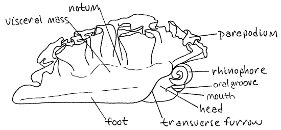
Tridachia crispata is a large sluglike opisthobranch, usually at least 5 cm and sometimes as much as 10 cm in length. The body of a living individual is usually green with white spots but the pattern is variable. The body consists of a central elongate visceral mass with the foot below it and the head at its anterior end (Fig 1). The dorsal surface is the notum, or back. Tridachia has no mantle cavity, gill, or osphradium.
Laterally the notum bears a pair of broad, thin, winglike parapodia (Fig 1). The right and left parapodia join each other dorsal to the head. The lateral margins of the parapodia are much longer than the body length and consequently are folded or ruffled.
The long flat foot is on the ventral surface of the visceral mass. A shallow transverse furrow crosses the anterior end of the foot and separates the anterior portion of the foot from the posterior. The furrow deepens laterally where it separates the head from the visceral mass.
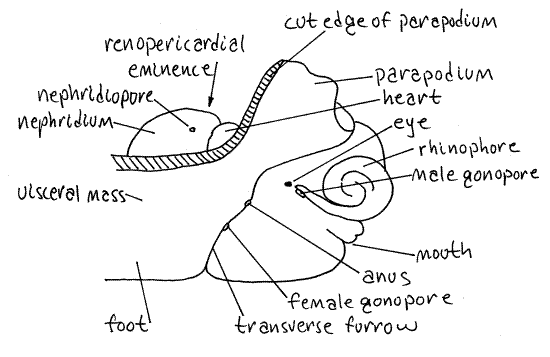
The prominent raised area on the midline of the anterior notum is the renopericardial eminence (Fig 2). The eminence is immediately posterior to the junction of the right and left parapodia. The posterior two thirds of this mound houses the nephridium, or kidney, and the nephridiopore is located on its right side.
The anterior third of the eminence contains the heart, whose rhythmic contractions are observable in living, unrelaxed specimens (Fig 2). Blood vessels from the parapodia can be seen entering the eminence. The largest are visible as two ridges running from the medial margins of the posterior parapodium to the eminence. Other shorter, less evident vessels drain the anterior regions of the parapodia into the heart. There is a total of six of these vessels, in three pairs.
Figure 1. Lateral view of a preserved Tridachia crispata. Redrawn from Marcus & Marcus (1960). Opist42L.gif

Figure 2. Lateral view of the right side of the head of a preserved specimen. Redrawn from Marcus & Marcus (1960). Opist43L.gif

The head sits dorsal to the anterior end of the foot. It is separated from the foot by a deep transverse oral groove (Fig 1). The mouth lies on the midline in this groove.
The head bears two thick tentacle-like rhinophores consisting of rolled extensions of the body wall. Each is tubular with a longitudinal rhinophoral slit along its lateral margin. A small black eye is located on the side of the head just posterior to the base of this slit.
There are several small, but biologically important, openings on the right side of the head (Fig 2). These pores are easily found in living specimens but are elusive in preserved material. They are best located while looking at the side of the head and then confirmed by probing gently with a minuten nadel (see Techniques chapter).
The male gonopore of these simultaneous hermaphrodites is at the proximal end of the rhinophoral slit on the right side of the head (Fig 2, 4). It is anterior to the eye and rhinophore.
The anus and female gonopore are both in the transverse furrow between the head and visceral mass (Fig 2, 4). The anus is the more dorsal of the two and is located in the groove near the base of the parapodium. The female pore is a little more ventrally situated but also lies in the transverse groove. Confirm the location of these pores with the nadel.
Internal Anatomy
" Make a shallow, median, dorsal incision with fine scissors beginning at the mouth and extending to the posterior end of the renopericardial eminence. Cut only the body wall and take care to avoid all internal structures. Pull the cut edges of the incision aside so you can see into the hemocoel, or body cavity.
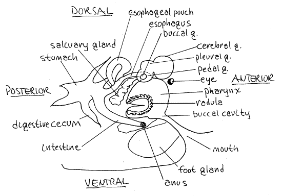
Fig 3. Internal anatomy of Tridachia crispata emphasizing the gut and nervous system. Redrawn from Marcus & Marcus (1960). Opist44La.gif

Hemal System
The pericardial cavity and the heart occupy the anterior third of the renopericardial eminence (Fig 2). Cut the membranous pericardium to open the pericardial cavity and reveal the heart within. The single, median, ovoid ventricle is conspicuous and easily seen once the pericardial cavity exposed.
The two atria are smaller and less easily found. Each is a thin-walled sac on a posterior lateral wall of the ventricle. A large, conspicuous, green (in life) aorta extends anteriorly from the ventricle and lies on the midline. It is the most conspicuous feature in this area. Trace the aorta anteriorly.
Nervous System
The conspicuous green portion of the aorta ends at the posterior border of the wide, white circumesophageal nerve ring. The ring is composed of several pairs of the usual molluscan ganglia including the pleural, buccal, pedal, and cerebral (Fig 3). Most evident are the two coalesced cerebral ganglia which form the dorsal portion of the ring. Several nerves can be seen extending laterally from the ring.
Excretory System
The nephridium, or kidney, occupies the posterior two thirds of the renopericardial eminence (Fig 2). Remove the body wall from the eminence and open the kidney to see its thick folded walls and inner lumen. It opens to the exterior via the nephridiopore found earlier.
Digestive System
Locate mouth on the ventral surface of the head. It opens into a small buccal cavity dorsal to itself (Fig 3, 12-42).
Immediately posterior to the buccal cavity is the large, white, bulbous pharynx. The pharynx is immediately anterior to the nerve ring. The muscles of the pharynx generate suction with which the snail sucks the juices from plants.
" The narrow, but muscular, esophagus exits the posterior end of the pharynx (Fig 3, 12-42). With fine scissors cut the nerve ring, aorta, heart and kidney along the dorsal midline to expose the esophagus ventral to them.
A single, hollow, spherical esophageal gland is located atop the middle region of the esophagus and opens into the esophagus (Fig 3). The gland is a blind pouch off the dorsal wall of the esophagus, a fact that can be demonstrated by opening the pouch.
A pair of intensely white, lobulated salivary glands lie lateral to the esophagus anterior to the esophageal gland (Fig 3). Their ducts pass through the nerve ring and empty into the posterior pharynx.
Posterior to the esophageal gland the esophagus continues the median stomach. The stomach is best found by opening the esophagus and following its lumen posteriorly through the digestive cecum (Fig 3). The walls of the stomach lumen are folded and appear ridged.
Two lateral digestive ceca connect with the stomach by ducts. The ducts divide repeatedly into numerous smaller tubelike diverticula in the surrounding tissues (Fig 3). The duct openings can be seen on the walls of the stomach and the ducts themselves are faintly visible in the translucent nearby tissue.
The intestine exits the stomach dorsally to run to the anus on the right. The intestine is white but is surrounded by green tissue. Use the nadel to trace it to the anus. Notice that the gut of Tridachia continues to show the effects of torsion. The anus is anterior, or the right side and the intestine runs anteriorly from the stomach to reach it.
" With fine scissors cut through the thick muscular folded dorsal wall of the pharynx to expose its lumen.
The radula lies on the floor of the pharynx where it can be seen as a narrow opaque, white, median line (Fig 3, 12-42). It consists of a single row of large teeth. The teeth are used to pierce the plant cells that are the food of these slugs so the pharynx can suck the juices from it. The tooth row curves around the anterior end of the floor of the pharynx. At the anterior, ventral end of the radula there is a radular sac, or ascus, that contains used and discarded teeth. The ascus is poorly developed in Tridachia.
>1a. Remove the opened pharynx and make a wetmount with it. With the compound microscope find the radula and its single tooth row. Find the ascus and its discarded teeth. <
Reproductive System
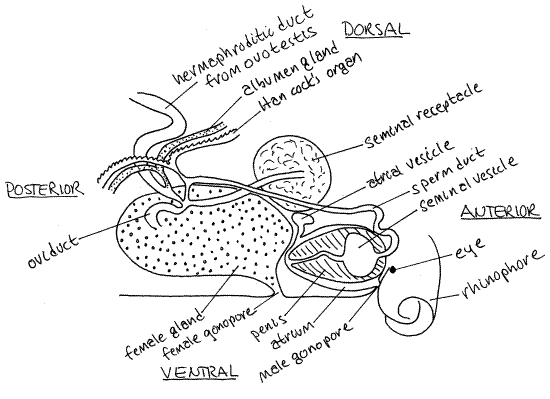
Sacoglossans are simultaneous hermaphrodites. Cross fertilization occurs with copulation and internal fertilization. The reproductive system is consequently complicated with much regional specialization of the gonoduct (Fig 4). The male and female gonopores have already been located on the right side of the head.
The hermaphroditic gonad, or ovotestis is composed of tubules scattered throughout the tissue of the notum and parapodia. These opaque, white ducts form an irregular network that is especially dense in the bases of the parapodia. Eggs and sperm exit the ovotestis via the hermaphroditic duct (Fig 4).
Figure 4. Hermaphroditic reproductive system of Tridachia crispata. Redrawn from Marcus & Marcus (1960). Opist45L.gif

Downstream from the ovotestis the hermaphroditic duct divides into an oviduct to the female organs and female pore and a sperm duct to the male organs and male pore.
The oviduct leads to a large white, opaque female gland to the right of the midline. This gland is elongate and hollow, with glandular walls. Distally the oviduct narrows and runs to the female pore. The walls of this terminal portion are thin and translucent. A bulbous seminal receptacle, for the storage of allosperm, is attached to the posterior (proximal) end of the female gland. It is also hollow and has thick, opaque, white walls like those of the female gland. The lumina of the receptacle and the female gland are continuous.
The sperm duct is the male branch of the hermaphroditic duct. It runs along the left side of the female gland to the spherical male atrium located in the tissue at the base of the right rhinophore. The sperm duct enters the atrium and opens into the seminal vesicle where autosperm are stored. The vesicle is located inside the base of the conical penis and the penis is in the atrium. An opening in the wall of the atrium leads to a very short duct that leads to the male gonopore.
" Open the atrium to find the penis and then open the base of the penis to find the oval seminal vesicle. "
During copulation the penis is everted through the male pore and inserted into the female gonopore of the partner. Spermatozoa from the seminal vesicle in the penis are deposited in the seminal receptacle of the partner. They will later be used to fertilize eggs passing through the female gland.
References
Hyman LH . 1967. The Invertebrates, Mollusca, vol. VI. McGraw-Hill, New York. 792p.
MacFarland FM. 1924. Expedition of the California Academy of Sciences to the Gulf of California in 1921. Opisthobranchiate Mollusca. Proc. California Acad. Sci. Ser. 4. vol 13 (25):389-420.
Marcus Ev, Marcus Er . 1960. Opisthobranchs from American Atlantic warm waters. Bull. Mar. Sci. Gulf, Caribbean. 10:129-203.
Ruppert EE, Fox RS, Barnes RB. 2004. Invertebrate Zoology, A functional evolutionary approach, 7 th ed. Brooks Cole Thomson, Belmont CA. 963 pp.
Supplies
Dissecting microscope
Small dissecting pan
Isotonic magnesium chloride
Seawater
#1 stainless steel insect pins
Dissecting set with microdissecting tools