Invertebrate Anatomy OnLine
Narceus americanus ©
Worm Millipede
29jun2006
Copyright 2001 by
Richard Fox
Lander University
Preface
This is one of many exercises available from Invertebrate Anatomy OnLine , an Internet laboratory manual for courses in Invertebrate Zoology. Additional exercises can be accessed by clicking on the links on the left. A glossary and chapters on supplies and laboratory techniques are also available. Terminology and phylogeny used in these exercises correspond to usage in the Invertebrate Zoology textbook by Ruppert, Fox, and Barnes (2004). Hyphenated figure callouts refer to figures in the textbook. Callouts that are not hyphenated refer to figures embedded in the exercise. The glossary includes terms from this textbook as well as the laboratory exercises.
Systematics
PanarthropodaSP, Arthropoda P, Mandibulata, Tracheata, Myriapoda SC, Progoneata,Dignatha, Diplopoda C, Chilognatha, Helminthomorpha, Eugnatha, Juliformia SO, Spirobolidae F(Fig 6-15, 20-14, 20-15)
Panarthropoda SP
Panarthropoda includes Onychophora, Tardigrada, and Arthropoda. These taxa share segmentation, a hemocoel, saccate nephridia, ecdysis of a secreted chitinous but non-collagenous exoskeleton, loss of locomotory cilia, a tubular, dorsal, ostiate heart in a pericardial sinus, a coelom reduced to end sacs and gonocoel, and paired segmental legs.
Arthropoda
Arthropoda, by far the largest and most diverse animal taxon, includes chelicerates, insects, myriapods, and crustaceans as well as many extinct taxa. The body is segmented and primitively bears a pair of jointed appendages on each segment. The epidermis secretes a complex cuticular exoskeleton which must be molted to permit increase in size. Extant arthropods exhibit regional specialization in the structure and function of segments and appendages. The body is typically divided into a head and trunk, of which the trunk is often itself divided into thorax and abdomen.
The gut consists of foregut, midgut, and hindgut and extends the length of the body from anterior mouth to posterior anus. Foregut and hindgut are epidermal invaginations, being derived from the embryonic stomodeum and proctodeum respectively, and are lined by cuticle, as are all epidermal surfaces. The midgut is endodermal and is responsible for most enzyme secretion, hydrolysis, and absorption.
The coelom is reduced to small spaces associated with the gonads and kidney. The functional body cavity is a spacious hemocoel divided by a horizontal diaphragm into a dorsal pericardial sinus and a much larger perivisceral sinus. Sometimes there is a small ventral perineural sinus surrounding the ventral nerve cord.
The hemal system includes a dorsal, contractile, tubular, ostiate heart that pumps blood to and from the hemocoel. Excretory organs vary with taxon and include Malpighian tubules, saccate nephridia, and nephrocytes. Respiratory organs also vary with taxon and include many types of gills, book lungs, and tracheae.
The nervous system consists of a dorsal, anterior brain of two or three pairs of ganglia, circumenteric connectives, and a paired ventral nerve cord with segmental ganglia and segmental peripheral nerves. Various degrees of condensation and cephalization are found in different taxa.
Development is derived with centrolecithal eggs and superficial cleavage. There is frequently a larva although development is direct in many. Juveniles pass through a series of instars separated by molts until reaching the adult size and reproductive condition. At this time molting and growth may cease or continue, depending on taxon.
Mandibulata
Mandibulata includes arthropods in which the third head segment bears a pair of mandibles. As currently conceived this taxon includes myriapods, hexapods, and crustaceans. Appendages may be uni- or biramous and habitats include marine, freshwater, terrestrial, and aerial.
Tracheata
Myriapods and hexapods share tracheae and a single pair of antennae and are sister taxa in Tracheata. Crustaceans, which have gills and lack tracheae, are excluded and form the sister group.
Myriapoda
The body consists of a head and trunk with numerous segments but no tagmosis into thorax and abdomen. Myriapoda includes the familiar centipedes and millipedes, as well as symphylans and pauropods.
Dignatha
This myriapod taxon comprises millipedes and pauropods, both of which have two pairs of mouthparts instead of the three of the ancestral tracheate, insects, centipedes, and symphylans. Millipedes have a pair of mandibles and a single gnathochilarium, which probably arose through fusion of the two first maxillae. The second maxillae are lost in dignathans.
Diplopoda
Most millipedes are elongate, often worm-shaped, mandibulates, although some are short and resemble woodlice. The body consists of a head and a trunk of many segments, most of which bear paired appendages. Most of the trunk segments occur in fused pairs, called diplosegments, each of which bears two pairs of legs and two pairs of spiracles externally. Internally each diplosegment has two pairs of ganglia, two pairs of ostia and two pairs of lateral blood arteries.
The head bears a pair of antennae, a pair of mandibles, and a pair of fused first maxillae. There is no second maxilla (= labium) and its segment is absent. The exoskeleton is strengthened with calcium salts and is correspondingly harder than that of most hexapods. The heart is an elongate ostiate dorsal tube extending most of the length of the body. Excretion is accomplished via a single pair of Malpighian tubules and a pair of maxillary glands. Respiration is via segmentally arranged tracheae with segmental spiracles.
The nervous system consists of a three-part mandibulate brain and a double, ganglionated, ventral longitudinal nerve cord. Millipedes are gonochoric with the gonopores between segments 2 and 3. Juveniles hatch with three pairs of legs and add body segments and additional legs with successive molts (Fig 20-12F).
Laboratory Specimens
Millipedes are either cylindrical in cross section (eg. Juliformia, Pentazonia, Fig 20-D, 20-9) or flattened dorsoventrally (eg. Polydesmida, Penicillata, Fig 20-8A,B). This account is written for a juliform millipede such as Narceus (formerly Spirobolus). Living millipedes can be anesthetized or killed by exposure to chloroform or ethyl acetate fumes, respectively. Living or preserved material can be used for this exercise.
External Anatomy
Place a living or preserved juliform millipede in a dry dish or small dissecting pan for study. Use a dissecting microscope to observe the specimen.
Juliform millipedes, such as Narceus and its relatives, have cylindrical bodies with well-developed heads (Fig 20-9). The body is circular in cross section and is divided into a shorthead and a long, multisegmented, leg-bearing trunk.
Head
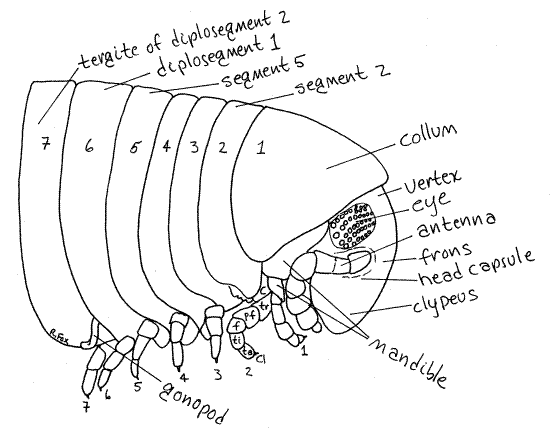
The dorsal and anterior surface of the head is covered by a heavily sclerotized exoskeletal plate known as the head capsule (Fig 1). The head is relatively small and is partly obscured by the enlarged tergite (collum) of the first trunk segment (Fig 1, 20-11).
The head capsule is toothed along its anterior, ventral border and comprises several regions (Fig 1). The dorsal posterior region, between the compound eyes, is the vertex. Anterior to the vertex, between the bases of the antennae, is the frons. Most of the remainder of the capsule is the clypeus (KLIP ee us), on the anterior-ventral surface. The labrum is the ventralmost extension of the clypeus (Fig 20-10A).
Each side of the capsule bears a lateral eye, consisting of a loose cluster of about 50 simple ommatidia. The ommatidia lack crystalline cones and these clusters are probably not homologous to the compound eyes of insects. The posterior sides of the head are formed by the basal article of the mandible, as you will soon see (Fig 1, 20-11).
Millipedes have three pairs of head appendages. The antennae are conspicuous structures arising from a pocket in the head capsule near the compound eyes (Fig 1). Each consists of seven articles. Note the groove in the head capsule into which the antenna fits when not in use.
The mouthparts, preoral cavity, and mouth are located on the ventral surface of the head. The preoral cavity is an external space from which the mouth opens. This space is enclosed and protected anteriorly by the clypeus and its ventral extension, the labrum. Laterally it is enclosed by the mandibles and posteriorly by the modified maxillae (gnathochilarium).
As dignathous myriapods, millipedes have only two pairs of mouthparts in contrast with the three pairs of most tracheates. The large, heavily sclerotized mandibles can be seen on either side of the posterior head (Fig 1, 20-10A). The two first maxillae are fused together on the midline to form a platelike structure known as the gnathochilarium (Fig 20-10A). The second maxillae have been lost in dignathans.
Use fine forceps to deflect the gnathochilarium posteriorly and find the massive mandibles hidden beneath the labrum. Note their toothed medial cutting surfaces. Each mandible consists of two articles. The basal, or proximal, article is visible on the side of the head. Try to push the mandibles aside so you can see the preoral cavity. The mouth opens from this space.
Figure 1. Head and anterior trunk of a male Narceus americanus from Balsam Grove, North Carolina. c = coxa, tr = trochanter, pf = prefemur, f = femur, ti = tibia, ta = tarsus, cl = claw. Milliped5La.gif
Trunk
The trunk of juliform millipedes has in excess of 50 rings, or annulations, but most are not simple segments. A few anterior rings correspond to individual body segments but most rings are composed of two fused segments and are called diplosegments (Fig 1, 2010B).
In these millipedes, the first five rings are true segments whereas all the remaining ones are diplosegments. In this account the term ring will be used in reference to any of the divisions of the trunk including segments and diplosegments.
Each ring is covered by a heavily sclerotized cuticular ring of exoskeleton. As in other arthropods the ring is composed of a dorsal tergite, two lateral pleurites, and a ventral sternite (Fig 20-10C).
Look at the cuticular ring of a typical diplosegment. Most of the ring is made of the large dorsal tergite (Fig 1, 20-10C). Small pleurites are fused with the lateral ends of the tergite to form a single horseshoe-shaped piece that covers the dorsal, lateral, and much of the ventral aspects of the ring. In juliforms, pleurites cannot be distinguished from tergites and the combined piece is usually referred to as the tergite. The small ventral gap in the ring is filled by the sternite, which articulates laterally with the tergite. The legs in turn articulate with the sternite (Fig 20-10B). Tergites are much larger than sternites, an arrangement that allows the animal to enroll into a coil. In flat millipedes such as the polydesmids, extensions of the tergites form eavelike lateral overhangs, the paranota, that are responsible for the characteristic flattened appearance of these millipedes (Fig 20-10C).
Successive cuticular rings are joined by thin, flexible, uncalcified articular membranes. Move two of the diplosegments apart and find the transparent articular membrane connecting them.
One or two pairs of spiracles open on the extreme medial ventral end of each tergite (Fig 20-10B). Segments have one pair of spiracles and diplosegments have two pairs.
The first two trunk segments are unlike all others. Both are segments and segment 1, which is immediately posterior to the head, bears a greatly enlarged tergite, known as the collum, over its dorsal and lateral surfaces (Fig 1, 20-9). This segment has no sternite articulated directly with the collum (1st tergite). The sternite is widely separated from the tergite and connected to it by a thin flexible articulating membrane (Fig 20-10A). The single pair of legs of this segment articulate with this sternite, as usual.
Segment 2 has a normal tergite but, like segment 1, its sternite is separated from it by an articulating membrane. The two ventral ends of the second tergite form two pointed ventral spines that point medially. Only the second segment has such spines. The second sternite is associated with the second pair of legs.
The remaining rings bear normal tergites and sternites. The first five rings are segments and each bears a single pair of legs. Rings posterior to the fifth are diplosegments and have two pairs of legs each.
Examine a pair of legs. Each leg is composed of seven articles, or divisions. They are named, in order from proximal to distal; coxa, trochanter, prefemur, femur, tibia, tarsus, and claw (Fig 1). The legs are uniramous and articulate with the sternites via their coxae. The trochanter attaches to the lateral margin of the coxa so consequently the legs extend to the sides of the animal, not ventrally.
The gonopores of both sexes are located on the third segment. In males there is a single gonopore at the end of a median, muscular, retractable penis. The penis is located ventrally on the articulating membrane between the second and third sternites. It is usually completely retracted into a pocket in this membrane. The external opening of the pocket is a transverse slit in the membrane at the base of the second pair of legs.
Females have two gonopores, each located on an oval swellings known as a vulva (Fig 20-11, 12C,D). Each vulva is situated ventrolaterally between the second and third segments.
The male gonopods are located on the seventh ring (Fig 20-12A,B). This is a diplosegment whose two pairs of legs are both modified as gonopods. The seventh ring (second diplosegment) of males is the genital diplosegment. The seventh ring of females is unmodified and its legs are like those of other diplosegments.
The posteriormost ring is the preanal ring. It does not bear legs and is surrounded by an unbroken cuticular ring. This is the only such ring on the body. Two large, clamshell-like cuticular valves are located posterior to the preanal ring. They open laterally to reveal a spacious rectal chamber into which the anus opens. Feces is delivered to this chamber where it is formed into fecal pellets.
References
Buck JB, Keister ML . 1950. Spirobolus marginatus, pp. 462-475 in Brown FA. (ed) . Selected Invertebrate Types. Wiley, New York. 597p.
Kaestner A. 1967. Invertebrate Zoology, vol. II. Arthropod Relatives, Chelicerata, Myriapoda. Wiley-Interscience, New York. 472p.
Keeton WT. 1960. A taxonomic study of the millipede family Spirobolidae (Diplopoda:Spirobolida). Mem. American Entomol. Soc. 17:1-146, pls. 1-18.
Ruppert EE, Fox RS, Barnes RB. 2004. Invertebrate Zoology, A functional evolutionary approach, 7 th ed. Brooks Cole Thomson, Belmont CA. 963 pp.
Shear WA . 1999. Millipedes. American Scientist, 87:232-239.
Supplies
Dissecting microscope
Small dissecting pan
Living or preserved juliform millipede
Ethyl acetate or chloroform for living specimens