Invertebrate Anatomy OnLine
Ecteinascidia ©
Orange tunicate
24may2007
Copyright 2005 by
Richard Fox
Lander University
Preface
This is one of many exercises available from Invertebrate Anatomy OnLine , an Internet laboratory manual for courses in Invertebrate Zoology. Additional exercises, a glossary, and chapters on supplies and laboratory techniques are also available at this site. Terminology and phylogeny used in these exercises correspond to usage in the Invertebrate Zoology textbook by Ruppert, Fox, and Barnes (2004). Hyphenated figure callouts refer to figures in the textbook. Callouts that are not hyphenated refer to figures embedded in the exercise. The glossary includes terms from this textbook as well as the laboratory exercises.
Systematics
Chordata P, Urochordata sP, Ascidiacea C, Enterogona O, Phlebobranchia sO, Perophoridae F
Chordata P
Chordata is characterized by a suite of apomorphies including a dorsal hollow nerve cord, notochord, pharyngeal gill slits, and a post anal tail (Fig 29-1). The ancestor was a fishlike deuterostome that swam using alternating contractions of right and left longitudinal axial muscles to create undulations of the body. The flexible, incompressible notochord prevented these contractions from compressing the body while allowing lateral deflection. The chordate central nervous system is a hollow, median, longitudinal nerve cord formed in the embryo by an invagination of surface ectoderm whose original function was probably sensory reception. Paired pharyngeal gill slits connect the lumen of the pharynx with the exterior and originally functioned in suspension feeding with respiration being added later. A muscular tail posterior to the anus is, although commonplace in chordates, an unusual feature not found in other taxa. It is an extension of the axial musculature and is the chief locomotory organ. An additional apomorphy is the endostyle, a region of pharyngeal endoderm, that secretes iodated compounds, either mucus or hormones.
Tunicata (= Urochordata) sP
Tunicates are highly derived and less like the ancestral chordates than are cephalochordates or vertebrates. At some time in the life cycle all possess a notochord, dorsal hollow nerve cord, pharyngeal gill slits, postanal tail, and endostyle but only the gill slits and endostyle are present in adults. Adult tunicates use the pharyngeal gill slits for suspension feeding. The larva is much more chordate-like than the adult and resembles a tadpole or fish, has all the chordate apomorphies, and is known as the tadpole larva. Metanephridia are absent and coelom is reduced to a pericardial cavity and gonads. As in cephalochordates the gut is dominated by an enormous pharynx surrounded by a water-filled atrium but, unlike cephalochordates, it is U-shaped with the mouth and anus anterior. Tunicates may be benthic or planktonic and solitary or colonial. All are marine.
Tunicata is traditionally divided into Ascidiacea (the benthic sea squirts in three taxa; Aplousobranchia, Phlebobranchia, and Stolidobranchia), Thaliacea (the pelagic salps, pyrosomes, and doliolids), and Appendicularia (the pelagic larvaceans). Recent molecular evidence and reevaluation of morphological evidence, however, suggests that Ascidiacea is paraphyletic and Tunicata should be reorganized into three different higher taxa (Fig 29-32). In this reorganization Stolidobranchia would be one higher taxon. Phlebobranchia plus Thaliacea would be the second taxon. Aplousobranchia plus Appendicularia is the final tunicate taxon. For now, however, the traditional classification will be followed.
Ascidiacea C
Ascidiacea is usually taken as representative of Tunicata, at least for the purposes of introductory laboratory exercises. Ascidians, or sea squirts, are sessile filter feeders that, as adults, bear little resemblance to their chordate relatives. Ascidians have a living, external, cellular exoskeleton, or tunic, underlain by epidermis. The tunic resembles connective tissue, except it isoutside the epidermis, and consists of cells, a secreted extracellular matrix, and ground substance. Much of it is a cellulose-like polysaccharide. In many ascidians blood vessels cross the epidermis to enter the tunic, a feature found in no other animal.
The gut is U-shaped and both openings are anterior, with the anus dorsal to the mouth. The gut is dominated by an enormous pharynx whose wall is perforated by numerous tiny gill slits. The pharynx is surrounded by a water-filled atrium into which the gill slits open and which itself opens to the sea. It is both respiratory organ and filter-feeding device. Water and food particles enter the pharynx and the water passes through the gill slits to the atrium and then out the siphon. Food, entangled in mucus secreted by the endostyle, remains in the gut and passes posteriorly to be digested.
The hemal system includes a heart, vessels, and blood spaces in the connective tissue. The heart is enclosed in a pericardial cavity derived from the ancestral coelom. The pattern of blood flow resembles that of the cephalochordates and early vertebrates except that the heart reverses direction periodically and the blood thus flows in both directions through the system. Ascidians have no structure recognizable as a kidney.
Ascidians are simultaneous hermaphrodites and the gonoducts open into the atrium. Some ascidians are solitary and may be relatively large. Others are colonial with tiny individual zooids in a common tunic.
Enterogona O
The gonads are in the gut loop and the neural gland is below the cerebral ganglion.
Phlebobranchia sO
Phlebobranchs have no postabdomen and may be solitary or colonial. The pharynx wall has raised longitudinal and transverse blood vessels but is not pleated. The inner surface of the pharynx bears projecting papillae that support the mucous feeding net. Papillae on the inner wall of the pharynx help hold the mucous net in place. No postabdomen is present. The tail of the tadpole is vertical in all except Perophora.
Biology
Ecteinascidia is a colonial, tropical sea squirt whose individual zooids reach lengths of about 2 cm. Colonies consist of clumps of closely spaced zooids with elongate posterior pedicles arising from a common stolon. Each zooid has its own tunic and complete complement of organs but blood vessels in the stolon maintain communication between zooids. The tunic is transparent and there is relatively little body wall musculature, making these animals ideal for study without dissection. Most of the internal anatomy is visible through the tunic. Variable amounts of orange pigment occur in the mantle and is clearly visible externally in living specimens. The orange color is lost in preserved material. Ecteinascidia retains and gestates its orange eggs in the oviduct and atrium and releases tadpole larvae from the atrium. Developmental stages, including the tadpole, are bright orange.
Laboratory Specimens
Living specimens can be collected on tropical or subtropical coasts or purchased from biological supply companies located in such areas. Ecteinascidia is available commercially as stained wholemount slides and this exercise is based on such slides. Commercially prepared slides each have a single zooid (Fig 1). At about 2 cm Ecteinascidia zooids are larger than is usual for compound ascidians making it large enough to see most of its structures without difficulty. .
If living specimens are available, water circulation and feeding should be observed through the transparent tunic using seawater tinted with non-toxic dyes (food coloring) or laden with suspended particles (carmine, India ink). For observation of smaller, less visible structures such as the beating heart and developmental stages the zooid can be easily removed from the tunic using fine forceps and iridectomy scissors.
Anatomy
Examine a wholemount of Ecteinascidia using 20X of the dissecting microscope. Ecteinascidia is a colonial ascidian in which individual zooids, each with a complete complement of viscera, are connected by a creeping stolon (Fig 29-12B). The zooid is elongate with a long tapered pedicle which connects with the horizontal stolon. The stolon may not be represented on the slide. A blood vessel extends from the zooid through the pedicle to the stolon. The zooid on the wholemount has been separated from the other zooids of its colony. It is probably distorted due to contraction during the fixation process and because it is severely depressed by the coverslip.
The wide blunt end of the zooid is anterior and the tapered end is posterior (Fig 1). Specimens on wholemounts are usually mounted so you are viewing either the right or left side. Be careful in the instructions that follow to distinguish between the animal’s right and left and directions on the slide. The ventral aspect is best recognized by finding a very long bright red (stained) groove, the endostyle. The endostyle begins near the buccal siphon and extends posteriorly for the length of the pharynx. The opposite edge, with the atrial siphon and rectum is dorsal.
Figure 1. A commercially prepared wholemount of a stained Ecteinascidia zooid viewed from its left side. Uro98L.gif
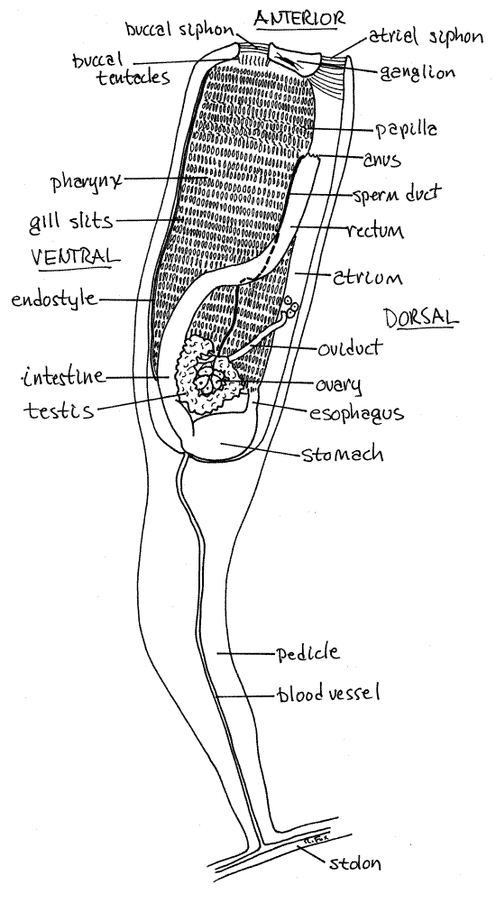
Before proceeding find the above landmarks and determine if you are looking at your specimen’s right or left side. As always, directions refer to the animal’s right and left, not necessarily yours. It is best to conduct most of your study with the left side uppermost, facing you. You can turn the slide over if necessary but be careful you don’t damage the coverslip. To be safe, suspend the slide (upside down) from two applicator sticks, one at each end of the slide, so the coverslip does not touch the stage.
The zooid is elongate with the anterior end (thorax) wider than the tapering posterior pedicle. (Distortion of the tunic may cause the posterior end to be wider than the anterior.) The transparent tunic is lightly stained (usually green) and surrounds the zooid. At the anterior end find the two siphons. The buccal siphon is near the center of the anterior end and the atrial siphon is dorsal to it (Fig 1). The opening of both siphons is flush with the truncate anterior end of the thorax but may not appear so in wholemount slides. If the specimen is distorted the siphons may not be at the tip of the body but posterior to it.
The buccal siphon is the anterior opening of the digestive system. The mouth lies just inside the siphon and is encircled by a ring of buccal tentacles, but these may be difficult to find in wholemounts. The buccal siphon and mouth open into the pharynx, which is the region of the gill slits and the site of filtration. The pharynx is surrounded by a water jacket, the atrium which empties to the exterior through the atrial siphon. The atrium receives seawater that exits the pharynx through the gill slits, feces from the gut, and gametes from the hermaphroditic gonads. In many species, including Ecteinascidia, it also functions as a gestation chamber.
The enormous, pale pink (stained) pharynx occupies most of the thorax. It is, of course, the anterior region of the gut into which the buccal siphon opens. It is adapted for filter feeding and its walls are perforated by abundant small oval gill slits. In most specimens about 30 rows of gill slits, with about 60 slits per row, are present. You are looking through both sides of the pharynx so you will see gill slits on its right and left sides superimposed on each other in a confusing jumble. Examine the pharynx with 100X of the compound microscope to see the gill slits more clearly but do not use high power on these thick slides.
Return the slide to the dissecting microscope and 20X. The endostyle is a long, conspicuous, red-staining groove extending the length of the ventral edge of the pharynx. It will be a narrow pale stripe flanked on each side by a thick heavily stained stripe. This appearance is due to the endostyle being a groove flanked by two thick ridges of tissue (Fig 29-15C).
The esophagus exits the pharynx dorsally at the posterior end (Fig 1). It is a short curved tube. When viewed from the right, the esophagus exits the dorsal posterior corner of the pharynx. The esophagus extends to the left and joins the stomach, which is a large chamber on the posterior margin of the pharynx. It extends from dorsal to ventral across the posterior end of the pharynx. Upon reaching the ventral edge of the zooid it narrows and curves anteriorly to pass diagonally to the left of the pharynx as the intestine. The transition from stomach is the pylorus and here the gut receives a duct from the digestive ceca. The ceca are not usually visible in these preparations. The intestine extends anteriorly then angles obliquely across the left side of the pharynx and becomes the rectum. The rectum extends straight anteriorly on the dorsal margin of the pharynx (Fig. 1). The appearance of the intestine and rectum vary depending on their contents, if any. They may be yellowish, or dark, or pale pink (if empty). The yellow color is due to the contents. Its walls can be seen to stain pink like the other regions of the gut. The rectum terminates at the anus beside the atrial siphon at the anterior end.
The anus empties into the atrium, which encloses the right, left, and dorsal sides of the pharynx. It can be seen along the dorsal margin of the pharynx, between the pharynx and the tunic.
Look in the loop between the esophagus and the intestine, on the left of the animal, for the gonads. The testis in a horseshoe shaped, lobed, intensely stained, red circle in this loop. A distinct dark red sperm duct extends from one end of the testis to the atrium where it opens near the anus. The yellow ovary is a cluster of large eggs located in the center of the horseshoe of the testis. The oviduct extends from the ovary to the atrium but it does not follow the sperm duct. Instead it reaches the atrium at a more posterior position and extends over the dorsal margin of the pharynx to opens into the right side of the atrium (Fig 1). The oviduct is difficult to see unless it contains embryos being gestated.
The heart, cerebral ganglion, neural gland, and digestive ceca are not usually visible in these wholemounts.
References
Berrill NJ . 1950. The Tunicata, with an account of the British Species. Ray Society, 133: 1-354.
Ruppert EE, Fox RS. 1988. Seashore animals of the Southeast. Univ. South Carolina Press, Columbia. 429pp.
Ruppert EE, Fox RS, Barnes RB. 2004. Invertebrate Zoology, A functional evolutionary approach, 7 th ed. Brooks Cole Thomson, Belmont CA. 963 pp.
Van Name WG. 1921. Ascidians of the West Indian Region and the Southeastern United States. Bull. Am. Mus. Nat. Hist., 44(16): 283-494.
Van Name WG. 1945. The North and South American ascidians. Bull. Am. Mus. Nat. Hist., 85:1-476, pls 1-31.
Supplies
Ecteinascidia living, preserved, or wholemounts
Wholemount slides are available from Carolina Biological
( www.carolina.com )
Living specimens from Gulf Specimen Marine Laboratories Inc ( www.gulfspecimen.org ).
Compound and dissecting microscopes