Invertebrate Anatomy OnLine
Girardia tigrina ©
Brown Planarian
28may2007
Copyright 2001 by
Richard Fox
Lander University
Preface
This is one of many exercises available from Invertebrate Anatomy OnLine , an Internet laboratory manual for courses in Invertebrate Zoology. Additional exercises can be accessed by clicking on the links on the left. A glossary and chapters on supplies and laboratory techniques are also available. Terminology and phylogeny used in these exercises correspond to usage in the Invertebrate Zoology textbook by Ruppert, Fox, and Barnes (2004). Hyphenated figure callouts refer to figures in the textbook. Callouts that are not hyphenated refer to figures embedded in the exercise. The glossary includes terms from this textbook as well as the laboratory exercises.
Systematics
Bilateria, Protostomia, Platyhelminthes P, Turbellaria C, Seriata, Tricladida O, Dugesiidae F (Fig 10-32)
Platyhelminthes P
Flatworms, or platyhelminths, are bilaterally symmetrical metazoans with three tissue layers. Unlike most triploblastic animals, they are compact and have no coelom (body cavity) surrounding the viscera and no hemal system. The gut, if present, has a single opening to the exterior. An anterior brain with associated concentration of sense organs is present, as expected of bilaterians. Flatworms are complex animals with elaborate hermaphroditic reproductive systems. Fertilization is internal with copulation. They may be free-living or parasitic.
Turbellaria C
The 4500 described species of free-living flatworms are included in the heterogeneous taxon Turbellaria (Fig 10-32*), whereas the parasitic flukes and tapeworms belong to another taxon, Neodermata. Turbellarians are carnivores varying in length from less than 1 mm to as many as 60 cm in length but the great majority are small. Many are roughly the size of ciliate protozoans, with which they are easily confused. The body is cylindrical in small species (microturbellaria, Fig 10-2) and dorsoventrally flattened in large (macroturbellaria, Fig 10-1). The epidermis is ciliated. The mouth is located somewhere on the ventral midline and opens into a blind gut, or gastrovascular cavity, which lacks an anus and sometimes lacks a lumen. Most are aquatic, inhabiting the oceans and fresh water, but a few are found in moist terrestrial environments. Most are benthic.
Osmoregulation and fluid regulation areaccomplished with protonephridia (Fig 10-20). There is no hemal system and transport is by diffusion, which is facilitated by small body size and flattening. Nutriment is delivered to tissues by diffusion from the gut, whereas oxygen diffuses across the body surface.
The end product of nitrogen metabolism is ammonia, which is lost by diffusion across the body surface. The nervous system consists of a bilobed brain, or cerebral ganglion, from which longitudinal nerve cords arise and extend posteriorly for the length of the body (Fig 10-11). Light-sensitive pigment-cup ocelli occur in various positions on the body. Turbellarians are hermaphroditic with internal fertilization.
Tricladida O
Because of the great heterogeneity of Turbellaria there is no typical example although the triclad, Girardia tigrina (= Dugesia tigrina), is often used in teaching laboratories as if it were. Triclads are macroturbellaria in which the gut comprises three branches, or rami (Fig 1,10-14D). One ramus is anterior and two are posterior to the plicate pharynx situated ventrally, near midbody, at the junction of the three rami. Each ramus is itself branched with many ceca ramifying to reach and deliver food throughout the body
Laboratory Specimens
Girardia tigrina is one of several species of freshwater triclad flatworms collectively known as "planarians". This exercise employs commercially available wholemounts, cross sections, and living cultures. The species supplied by commercial houses vary but are similar and all are suitable for this exercise. The exercise is written specifically for "brown planaria".
Triclads inhabit freshwater where they are most common under stones, leaves, or other objects near the shore. They are negatively phototactic. They can be collected in local fresh waters, if present, by placing small pieces of lean beef on the bottom for 10-15 minutes.
Wholemount Slides
With the dissecting microscope or low power of the compound microscope study a composite wholemount with two fixed and stained planarians, one with the gut injected with dye and the other plain, with uninjected gut (Fig 1, 10-31*). Note the dorsoventral flattening and bilateral symmetry. Locate the anterior-posterior axis, which is the axis of symmetry. The plane of symmetry includes this axis and divides the worm into right and left sides. Specimens are usually mounted on the slide with the back, or dorsal surface, up.
The anterior end of the body is the head and the remainder is the trunk. Girardia tigrina has a pointed triangular head but the head of some species is blunt and rounded. The posterior limit of the head is marked by a pair of lateral, ciliated, chemosensory protrusions, the auricles. These are variously developed in different species and are sometimes absent. The two darkocelli, or eyespots, are easily seen at the level of the auricles.
The digestive system is best studied using a specimen that has been fed a colored substance such as carmine powder or carbon black and thus has a gut filled with pigment. A wholemount of such a specimen should be available, either on the slide you are now using or mounted separately.
Locate the cylindrical pharynx lying on the midline at the center of the body (Fig 1, 10-31A). The pharynx is a muscular, protrusible tube housed in a spacious pharyngeal cavity which opens to the exterior via the mouth. The narrow pharyngeal lumen, which may contain pigment, occupies the center of the pharynx. The pharyngeal cavity is enclosed by the pharyngeal sheath.
Figure 1. The planarian, Girardia tigrina, in dorsal view. Flatworm27L.gif
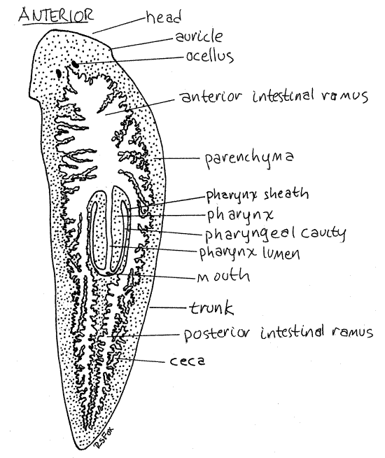
The mouth, which is rarely visible on wholemounts, is a small pore on the ventral midline of the body immediately posterior to the pharynx. It is the opening of the pharyngeal cavity to the exterior. During feeding, the pharynx lengthens to many times its resting length and is extended out of the mouth to reach the food.
The proximal end of the pharynx opens into the intestine which, in triclads, immediately divides into one anterior and two posterior rami. The name Tricladida (clad = branch) alludes to the presence of the three rami. Each ramus ends blindly and bears abundant ceca, or diverticula, so that no area of the body is beyond effective diffusion distance from the food source.
Numerous protonephridia are present but are not visible in these preparations. Sometimes some features of the reproductive system can be seen posterior to the pharynx but it is rare that enough can be discerned to make sense of it (Fig 10-31E). Study of the nervous system is also impractical (Fig 10-11C).
Cross-section Slides
Examine a prepared slide of planaria cross sections. Use a commercial slides with three cross sections taken at different levels along the worm. There is usually one through the anterior end of the body, one through the pharynx (Fig 3), and one through the posterior part of the worm (Fig 2). Slides are not uniform however, and do not necessarily conform to this ideal, nor can you rely on the order in which the sections are arranged on the slide.
Locate and identify each of the three sections on your slide. The pharyngeal section is usually easy to recognize (Fig 3). A good pharyngeal section shows the large, unmistakable pharynx as a hollow, red circle surrounded by a narrow white ring in the center of the section. The anterior and posterior sections have one to many irregular circles distributed through the parenchyma (Fig 2). These circles are the intestinal rami and their ceca (Fig 2).
Anterior or Posterior Cross-section
Begin with either the anterior or posterior section (Fig 2), i.e. one that does not pass through the pharynx. It makes little difference which you choose and you may find it profitable to use both but be certain you do not have the pharyngeal section. Be sure you can tell dorsal from ventral. The ventral surface is usually flatter than the arched dorsal surface (in most slides it will be down).
Figure 2. Cross section through the body of Girardia tigrina posterior to the pharynx. Flatworm28L.gif
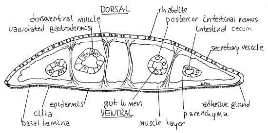
The body is covered by a monolayered, secretory epidermis (Fig 2). The ventral, but not the dorsal, epidermis is ciliated, a fact that can be verified by careful observation with high power (400X). The cilia are used for locomotion and the epithelial cells are multiciliated. The dorsal surface is not ciliated. Compare the dorsal and ventral epithelia to be sure you can recognize cilia. The epidermis is underlain by a distinct basal lamina, which is visible as a thin, dark line just inside the epidermis.
The dorsal epidermis contains numerous secretory vesicles and rod-shaped membrane enclosed secretions, the rhabdites (rhabd = rod). Rhabdites are synthesized by epidermal gland cells insunk (submerged) below the basal lamina into the parenchyma (Fig 10-5). When expelled at the surface, rhabdites absorb water and expand to become sticky mucus which may help trap small invertebrate prey.
Note the clusters of adhesive gland cells situated at the lateral edge of the ventral epidermis (Fig 2). These are part of a cilia-free adhesive zone that encircles the worm. These cells secrete an adhesive that helps the animal grip the substratum. The ventral epidermis bears numerous gland cells that secrete mucus.
A thick layer of body wall muscles, consisting of outer circular and inner longitudinal fibers, lies just inside the epidermis (Fig 2, 10-3C).
Inside the muscle layer, the interior of the worm is filled with a mesenchymal connective tissue, the parenchyma, consisting of cells in a fibrous extracellular matrix and having a loose, open appearance (Fig 10-13B). Many large epidermal gland cells are submerged into it but they nevertheless open to the surface via narrow necks passing through the basal lamina and epidermis.
Dorsoventral muscles can be seen passing vertically through the parenchyma connecting the muscle layers of the dorsal and ventral body walls. These muscles maintain the flat shape of the triclad body.
Scattered about in the interior are sections through the intestinal rami and their ceca (Fig 2, 10-13B). These are irregular circles of various sizes and unpredictable number. The clear space in the interior of each is the gut lumen and is surrounded by a monolayered epithelium of large, vacuolated cells, which may be secretory, absorptive, or phagocytic. This epithelium is usually referred to as the gastrodermis, or mucosal epithelium.
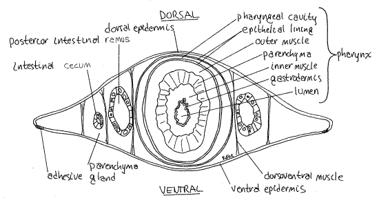
Pharyngeal Cross-section
Refer now to the pharyngeal cross section, which should resemble Figure 3. Review the now familiar features you identified on the other sections and then study the enormous pharynx in the center.
The pharynx occupies almost the entire center of the section and the body wall is very thin above and below it. Locate and identify the two white, unstained spaces associated with the pharynx. The one in the center of the pharynx is the pharyngeal lumen whereas that surrounding the pharynx is the pharyngeal cavity. This type of pharynx, known as a plicate pharynx and consisting of a muscular tube retractable into a sheath, is characteristic of triclads and polyclads (Fig 1, 10-16B).
Moving outward from the lumen in the center of the pharynx, the layers are, in order: pharyngeal lumen, gastrodermis, inner muscle layer, parenchyma (connective tissue), outer muscle layer, epithelium, pharyngeal cavity, and pharynx sheath epithelium (Fig 3).
Figure 3. Cross section through the pharyngeal region of Girardia tigrina. Flatworm29L.gif
Living Specimens
Place a living worm in a drop of water on a microscope slide without a coverslip. Living worms can be picked up with a plastic pipet but they must be ejected quickly or they will attach to its wall using their adhesive glands. They adhere tenaciously and are difficult to remove from the pipet when this happens.
Observe the worm with the dissecting microscope. Watch it as it moves across the slide. The major locomotory force is produced by the cilia of the ventral epidermis but muscular activity also plays a role in locomotion, especially in making turning movements. Manipulate the worm with a tiny needle to encourage it to change directions. Try to discover the contributions of the musculature to this maneuver and think about which muscles would be involved in making a turn. Triclads do not swim.
Push the worm gently with the needle and look for evidence of adhesive ability. Where do the adhesive cells seem to be located? _________________
Observe the animal with transmitted light and look for the intestine and its ceca. These may be obvious, especially if the worm has been fed recently.
> a. Position a coverslip over the worm and place the slide on the compound microscope. The weight of the coverslip will squeeze the worm enough to immobilize it and make it thin enough to see some internal structure. Remove some water from the preparation to further squeeze the animal. Do not use 400X on these slides.
Focus, with 100-200X, on the edge of the head, reduce the light, and look for evidence of beating cilia. Most of the animal's cilia are ventral and thus difficult to see in a wholemount but the head bears cilia associated with chemosensory receptors on the auricles and their activity is obvious. Look for them on the edge of the auricles. The name "turbellaria" means "little disturbance" and is a reference to the movement of water caused by the cilia of the auricles. Observe the cilia with phase contrast if you have it.
Increase the light so you can illuminate some of the interior. At 40X you may be able to see gut diverticula, especially if the animal has eaten recently. The pharynx is the conspicuous, long, pale area in the center of the body. If the animal is squeezed sufficiently, you may see the pharynx clearly and you may even see the mouth opening at its posterior end. The pharynx will probably move about in the pharyngeal chamber and may increase in length. <
> b. Place three or four worms in the center of a small (6 cm) culture dish. Place a small piece (about 2-3 mm in diameter) of hard-boiled egg yolk, lean beef, or beef liver in the dish with the worms. Turn the microscope and room lights off. Girardia tigrina is negatively phototactic and may unwilling to feed in bright light although sometimes it doesn’t seem to matter. Put the dish on the stage of the dissecting microscope being careful that you do not disturb the worm. If you are fortunate, you will see the worm protrude its pharynx and feed (Fig 10-31D). The sight is impressive and you may be startled by the length of the pharynx. Egg yolk is especially good for this exercise because the bright yellow yolk granules are easy to see as they move up the pharyngeal lumen to accumulate in the intestine. After several minutes of feeding look at the gut to see if the ceca and rami can now be visualized. <
Reproduction
Girardia tigrina typically reproduces asexually by architomy (Fig 10-22B), a type of fission in which the worm divides into two fragments without prior differentiation of new parts. Transverse cleavage just posterior to the pharynx divides the worm into an anterior, nearly normal, worm with head, mouth, pharynx and most of the gut, and an incomplete, headless posterior mass of tissues which must replace its missing parts.
Following division, the anterior end behaves normally but the posterior end remains immobile until regeneration is complete and the missing parts replaced. You are not likely to see fission occurring but, if your laboratory maintains populations in aquaria, it is quite possible that you will see these headless lumps stuck on the walls of the aquaria.
> c. Triclads have remarkable regenerative abilities and are capable of repairing damage or replacing missing body parts if experimentally fragmented. The process mimics architomy. Add about 20 ml of pond water to the dish containing your specimen(s). Remove the egg yolk and any worms in excess of one large worm.
Use a sharp scalpel to cut the remaining specimen in pieces. You decide how you want to cut the worm. Some obvious possibilities are to bisect it with transverse or longitudinal cuts. Or you could trisect it. Or you could split the head to see if you can produce a two-headed worm (if you choose this option you will need to visit the lab daily to renew the incision and keep the two halves of the head from growing back together.
Use a Sharpie to label the side of the dish with your name and the date. Cover the dish with another dish and set it aside in a dark area of the laboratory. Make careful sketches of the pieces of the worm. Observe your specimens during each laboratory period for the remainder of the semester. Make sketches each week and compare them with earlier sketches. Do you see evidence of regeneration? <
*Hyphenated call-outs, such as this one, refer to figures in Ruppert, Fox, and Barnes (2004). Those without hyphenation refer to figures embedded in this exercise.
References
Brown FA . (ed) 1950. Selected Invertebrate Types. Wiley, New York. 597p.
Hyman LH. 1951. The Invertebrates:Platyhelminthes and Rhynchocoela, vol. II. McGraw-Hill, New York. 550p.
Pennak RW . 1989. Fresh-water Invertebrates of the United States, 3 ed. Wiley, New York. 628p.
Petrunkevitch A . 1916. Morphology of Invertebrate Types. MacMillan, New York. 263p.
Pierce SK, Maugel TK. 1987. Illustrated invertebrate anatomy. Oxford Univ. Press, Oxford.
Rieger RM, et al. 1991. Platyhelminthes: Turbellaria, in Harrison, F. W. & B. J. Bogitsh (eds.). 1991. Microscopic Anatomy of Invertebrates vol. 3 Platyhelminthes and Nemertinea . Wiley-Liss, New York. 347p.
Riser NW, Morse MP (eds). 1974. Biology of the Turbellaria. McGraw-Hill, New York. 530 pp.
Ruppert EE, Fox RS, Barnes RB. 2004. Invertebrate Zoology, A functional evolutionary approach, 7 th ed. Brooks Cole Thomson, Belmont CA. 963 pp.
Supplies
Compound microscope
Dissecting microscope
Slides and cover glasses
Plastic Pasteur pipet
6 cm Carolina culture dish
Hard-boiled egg yolk
Prepared slides: stained wholemount with filled gut, stained cross sections of anterior, middle, and posterior regions,
Cultures: brown planaria
|
Item |
Source |
|
Composite wholemount slides (gut injected, gut plain) |
Carolina, Triarch, Ward’s |
|
Cross sections slides at three levels |
Carolina, Triarch, Ward’s |
|
Living Girardia tigrina (as Dugesia tigrina) |
Carolina, Ward’s |