Invertebrate Anatomy OnLine
Cephalobus ©
Roundworm
5jul2006
Copyright 2001 by
Richard Fox
Lander University
Preface
This is one of many exercises available from Invertebrate Anatomy OnLine , an Internet laboratory manual for courses in Invertebrate Zoology. Additional exercises, a glossary, and chapters on supplies and laboratory techniques are also available at this site. Terminology and phylogeny used in these exercises correspond to usage in the Invertebrate Zoology textbook by Ruppert, Fox, and Barnes (2004). Hyphenated figure callouts refer to figures in the textbook. Callouts that are not hyphenated refer to figures embedded in the exercise. The glossary includes terms from this textbook as well as the laboratory exercises.
Systematics
Cycloneuralia SP, Introverta, Nematoida, Nematoda P, Secernentea C, Rhabditida O, Cephalobina sO, Cephaloboidea SF, Cephalobidae F, (22-35, 9-26)
Cycloneuralia SP
Cycloneuralia includes Gastrotricha, Nematoda, Nematomorpha, Priapulida, Kinorhyncha, and Loricifera. Most have a secreted cuticle and lack locomotory cilia so that locomotion is accomplished with muscles. The brain is a circular band around the anterior gut composed of forebrain, midbrain, and hindbrain (Fig 22-2A). The pharynx is radially symmetrical (Fig 22-15). In most the body is compact without a body cavity but in large species there may be a spacious hemocoel. Eutely, with about 1000 cells, is common.
Nematoda
The 20,000 known nematode species inhabit terrestrial, marine, and freshwater environments and are found in almost all moist habitats. The taxon includes numerous plant and animal parasites, many of which are of medical or agricultural importance, but most are free-living (non-parasitic). Most nematodes, or roundworms, are long, slender, almost featureless externally, tapered at both ends, and round in cross section (Fig 22-7A). The body cavity, if present, is a hemocoel derived from the blastocoel.
The body is covered with a thick extracellular cuticle secreted by a cellular or syncytial epidermis that is molted during juvenile development (Fig 22-11, 22-9). The epidermal nuclei are sunken below the epithelial layer into four longitudinal epidermal cords that extend the length of the animal. The body wall has well-developed longitudinal but no circular muscles.
The gut is complete with terminal anterior mouth and subterminal posterior anus. It comprises ectodermal foregut and hindgut and an endodermal midgut. The body cavity, or hemocoel, is not lined with mesothelium and there is no muscle, connective tissue, or other mesodermal derivative associated with the midgut.
The nervous system is a ganglionated circumenteric ring, hence the name “cycloneuralia”, with several longitudinal nerve cords, the most important of which is the ganglionated, double, ventral cord (Fig 22-11A). The nerve cords are located in the longitudinal epidermal cords, along with the epidermal nuclei. Cytoplasmic innervation processes from the longitudinal muscles extend to the longitudinal nerve cords and serve the function of motor neurons, which are absent. Sensory equipment may include unique chemosensory amphids and sensory bristles around the mouth.
Most nematodes lack cilia or flagella, even in the sperm. There are, however, ciliary derivatives in the amphids (Fig 22-8B) and cilia are present in the gut epithelium of some nematodes (Fig 22-16A). Roundworms are ammonotelic and nitrogen excretion is mostly by diffusion across the body wall. Osmoregulation is accomplished by an excretory canal system in some and perhaps by excretory glands. No nephridia are present.
Nematodes are typically gonochoric and fertilization is internal with copulation. Sexual dimorphism is common. Nematode sperm have no flagella and probably employ amoeboid locomotion. Nematode development features a phenomenon known as chromosome diminution in which much of the chromosome material of presumptive somatic cells degenerates and is lost (Fig 22-20). Germ cells, however, retain the full complement of genetic material. Development is direct and includes four juvenile and one adult instar separated from each other by molts. Most nematodes are small (<3 mm) and free-living but some of the parasitic species, such as Ascaris, may reach 50 cm.
Secernentea C
Once known as Phasmidia, Secernentea includes terrestrial nematodes and many important parasites. Free-living, non-parasitic species, such as Cephalobus, are usually soil dwellers. Phasmids are present and the amphids are porelike. Excretory canals, and sometimes excretory glands, are present (Fig 22-17). Epidermal cells can be mono- or multinucleated.
Laboratory Specimens
Cephalobus is a small, free-living nematode that inhabits soil where it feeds on dead plant material and bacteria. It is viviparous and females gestate the embryos and release juveniles. Living cultures are available at low cost from biological supply companies.
Behavior
Place a small bit of culture in poikilotherm saline solution (see Supplies chapter) in a small culture dish and observe the worms with the dissecting microscope. Adults are 1 mm or less in length and exhibit the quintessential vermiform shape typical of nematodes, being cylindrical in cross section and tapered at both ends. The anterior end, however is blunter than the acutely pointed posterior end and the two are easily distinguished.
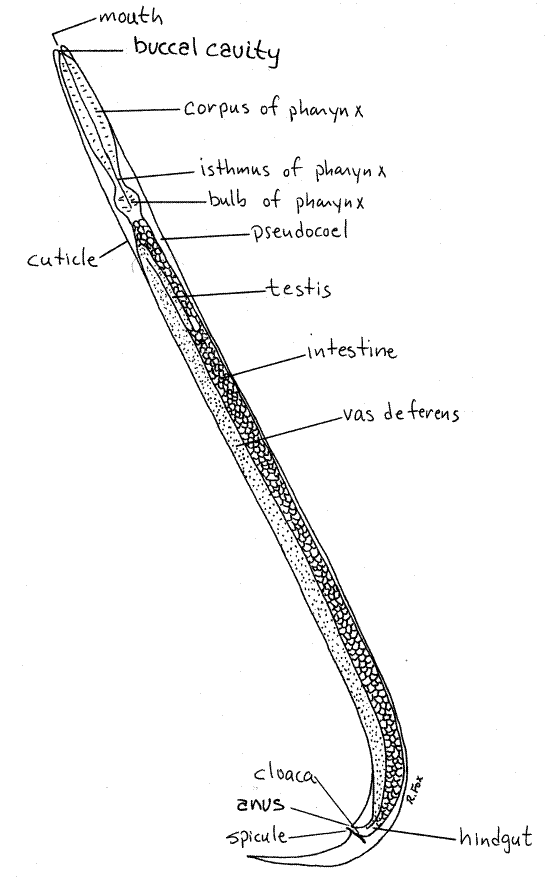
Figure 1. Male Cephalobus. Nematode41La.gif

Note the characteristic thrashing motion that results from the alternating contraction of dorsal and ventral longitudinal body wall muscles in the absence of circular muscles (Fig 22-12A). This motion is ineffectual in a smooth-bottom culture dish but in a complex substratum, with sand grains or other particles to push against, the worms move rapidly and efficiently in the desired direction.
>1a. Add a few sand grains to the dish and observe the efficacy of the longitudinal muscles in a complex substratum. <
Anatomy
Make a wetmount of a few nematodes and cover it with a coverslip. Do not use Protoslo or wax feet on the coverslip. Examine the preparation with the compound microscope and find relatively inactive worms to study. It may be necessary to heat the slide slightly to kill the worms so you can observe them. This is accomplished with a small flame from a butane lighter or small gas laboratory burner. Hold the flame briefly below the slide. More than one attempt may be required to achieve the correct amount of heat.
Search the slide for specimens and distinguish between males and females. Choose the largest specimens you can find for observation. The male posterior end tapers quickly to a point and is curved whereas that of females tapers more gradually (Fig 1, 2). Males also have a pair of copulatory spicules protruding from the cloaca near the posterior end of the worm. Females, being viviparous, usually contain juvenile worms of various sizes (Fig. 2). Study representatives of both sexes.
In both sexes the reproductive system is a single tube extending anteriorly from the gonopore but the location of the gonopore differs. The upper, anterior end of the tube is reflexed, or bent posteriorly, and is the gonad, where gametogenesis begins and gonial cells are produced (Fig 1, 2, 22-10D). Downstream portions are modified for various functions in the two sexes.
The integument consists of a thick cuticle, which is underlain by the epidermis, and a thick layer of longitudinal muscles (Fig 22-10B). There are no circular muscles and no mesothelium. The body cavity is an unlined hemocoel filled with the digestive and reproductive systems.
The gut and reproductive systems occupy all of the space in the hemocoel and each is likely to obscure your view of the other. You should work at 400X and use the fine focus to take optical sections in an attempt to resolve the parts of the two systems. The anterior end of the gut is easy to see and distinguish because there is no reproductive system in this part of the worm.
Male
Find a male and study it first (Fig. 1). Distinguish between the anterior and posterior ends. The anterior end tapers to a blunt point whereas the posterior end tapers abruptly to a sharp tip. The gut is the most conspicuous feature. It begins with a terminal mouth at the extreme anterior end of the worm. The mouth opens into a small buccal cavity (Fig 1, 22-13A). The buccal cavity is followed by the large pharynx that is easy to see because of its thick muscular walls (Fig 1, 22-14B). The pharynx is part of the foregut and as such is ectodermal and lined with cuticle. The thickness of its walls is due to radial muscles which, upon contraction increase the diameter of the lumen thus creating a suction that draws food into the mouth (Fig 22-15). The pharynx has three distinct parts. The anterior region is the corpus and is the longest (Fig 1). The corpus is followed by a narrow isthmus which opens into a spherical pharyngeal bulb. The brain is a band around the pharynx.
The midgut, or intestine, which is endodermal and is not lined by cuticle, follows the pharyngeal bulb and extends as a thick tube posteriorly almost to the posterior end of the worm (Fig 22-10A). At about the level at which the body begins to taper abruptly, the intestine narrows and turns ventrally to become the cloaca (Fig 22-18A). The cloaca opens to the exterior via theanus on the ventral midline.
The male reproductive system consists of a single long tube extending over most of the length of the worm (Fig 1, 22-10D, 22-18A). It is reflexed at the anterior end although this is not always apparent. The reflexed anterior end of the tube is the testis, where primordial germ cells undergo mitotic divisions to produce spermatogonia. The remainder of the tube is the vas deferens, or sperm duct, which stores sperm and joins the hindgut. The vas deferens is regionally specialized into a seminal vesicle for sperm storage, a secretory prostate, and a muscular ejaculatory duct. The chamber formed by the union of the vas deferens and the hindgut is the cloaca. The cloaca opens to the exterior by the anus. A pair of copulatory spicules may be visible beside, or protruding from, the cloaca.
Female
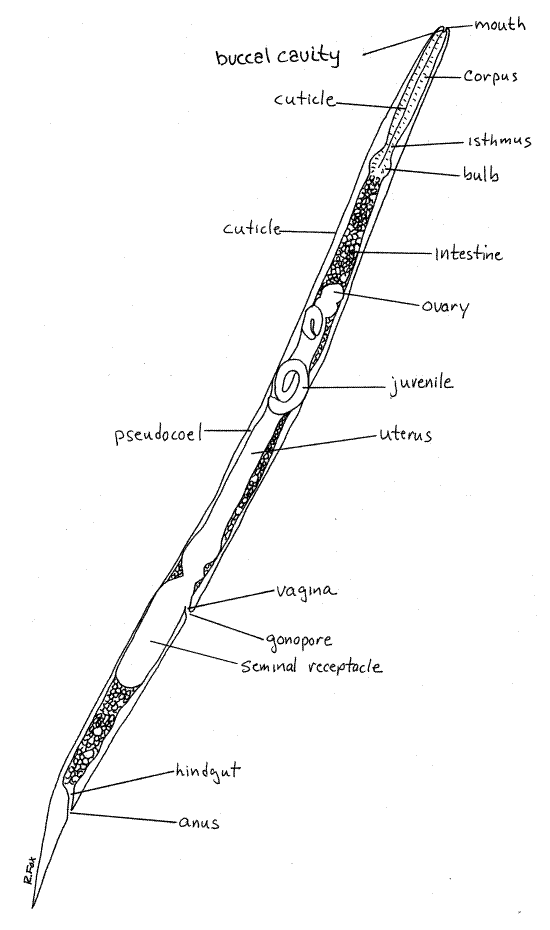
Now find a female and study it (Fig. 2). Distinguish between the anterior and posterior ends. The anterior end tapers to a blunt point whereas the posterior end tapers gently to a sharp tip. The easiest way to recognize females is by the presence of juvenile worms in the reproductive system (Fig 2). The hemocoel of females contains the gut and reproductive system, as does that of the male, but appears more crowded because the reproductive system is larger.
The female gut is similar to that of males except that there is no cloaca. In female nematodes the gut and reproductive system are independent of each other and they do not share ducts or openings as they do in the male. Consequently there are two external openings rather than one (Fig 2, 22-10A).
You should be able to find the same regions of the gut, with the exception of the cloaca, that you found in the male. The anterior mouth opens into a small buccal cavity which leads to the large, conspicuous pharynx (Fig 2). The pharynx is divided into corpus, isthmus and bulb. The brain is a band around the pharynx. The intestine, or midgut, extends posteriorly from the pharyngeal bulb as a large tube almost the same diameter as the body. It may be difficult to distinguish the intestine from the uterus. In most commercial cultures the gut contains recognizable potato granules that can be used to identify the gut. The intestine narrows to become the short hindgut, which then opens to the exterior via the anus on the ventral midline very close to the posterior end of the body. Females have no cloaca.
In Cephalobus the female reproductive system is a single large tube that may fill most of the hemocoel and obscure your view of the gut. The tube ends blindly at its anterior end and opens to the exterior via female gonopore, on the midventral line about 2/3 of the distance from the tip of the head to the tip of the tail. The tube is reflexed at the anterior end as is that of males. The upstream, anterior end is the ovary where oogenesis begins with production of oogonia from primordial germ cells. The ovary is difficult to discern in most specimens.
Figure 2. A female Cephalobus. Nematode42La.gif

Posteriorly, the ovary widens to become the much larger and more conspicuous uterus (Fig 2, 22-19). In most mature females you should see coiled juvenile worms of various ages in the uterus. The uterus narrows to become the short vagina which opens to the exterior via the female gonopore. The gonopore is on the midventral line about 2/3 of the distance from the anterior to the posterior end of the worm. The seminal receptacle is a large diverticulum of the vagina on the posterior side of the gonopore (Fig 2). It receives and stores sperm following copulation. In many nematodes the reproductive system consists of two twin tubes, each with ovary and uterus, which join to form the vagina (Fig 22-19).
During copulation the male inserts the copulatory spicules into the vagina to hold it open while sperm are ejaculated from the cloaca (Fig 22-18D). Sperm are stored in the seminal receptacle and make their way upstream to fertilize the oogonia as they leave the ovary. Once fertilized, the zygotes begin development and are gestated in the uterus until they become small worms. They are then released from the vagina through the gonopore.
References
Ho J-S . 1978. Laboratory Manual for Invertebrate Zoology Emphasizing Marine Forms. Hwong, Los Alamitos, Calif. 152p.
Hyman LH. 1951. The Invertebrates: Acanthocephala, Aschelminthes, and Entoprocta. The coelomate Bilateria, vol. III. McGraw-Hill, New York. 572pp.
Ruppert EE, Fox RS, Barnes RB. 2004. Invertebrate Zoology, A functional evolutionary approach, 7 th ed. Brooks Cole Thomson, Belmont CA. 963 pp.
Supplies
Dissecting microscope
8-cm culture dish
Compound microscope
Laboratory burner
Slides coverslips
Cephalobus culture
poikilotherm saline solution (0.5-0.7%)