Invertebrate Anatomy OnLine
Balanus eburneus ©
Acorn Barnacle
26jun2006
Copyright 2001 by
Richard Fox
Lander University
Preface
This is one of many exercises available from Invertebrate Anatomy OnLine , an Internet laboratory manual for courses in Invertebrate Zoology. Additional exercises can be accessed by clicking on the links on the left. A glossary and chapters on supplies and laboratory techniques are also available. Terminology and phylogeny used in these exercises correspond to usage in the Invertebrate Zoology textbook by Ruppert, Fox, and Barnes (2004). Hyphenated figure callouts refer to figures in the textbook. Callouts that are not hyphenated refer to figures embedded in the exercise. The glossary includes terms from this textbook as well as the laboratory exercises.
Systematics
Arthropoda P, Mandibulata, Crustacea sP, Eucrustacea, Maxillopoda SC, Progonomorpha, Thecostracomorpha, Thecostraca, Cirripedia C, Thoracica O, Balanomorpha sO, Balanoidea SF, Balanidae F, (Fig 16-15, 19-89, 19-90)
Arthropoda P
Arthropoda, by far the largest and most diverse animal taxon, includes chelicerates, insects, myriapods, and crustaceans as well as many extinct taxa such as Trilobitomorpha. The segmented body primitively bears a pair of jointed appendages on each segment. The epidermis secretes a complex cuticular exoskeleton which must be molted to permit increase in size. Extant arthropods exhibit regional specialization in the structure and function of segments and appendages but the ancestor probably had similar appendages on all segments. The body is typically divided into a head and trunk, of which the trunk is often further divided into thorax and abdomen.
The gut consists of foregut, midgut, and hindgut and extends the length of the body from anterior mouth to posterior anus. Foregut and hindgut are epidermal invaginations, being derived from the embryonic stomodeum and proctodeum respectively, and are lined by cuticle, as are all epidermal surfaces of arthropods. The midgut is endodermal and is responsible for most enzyme secretion, hydrolysis, and absorption.
The coelom is reduced to small spaces associated with the gonads and kidney. The functional body cavity is a spacious hemocoel divided by a horizontal diaphragm into a dorsal pericardial sinus and a much larger perivisceral sinus. Sometimes there is a small ventral perineural sinus surrounding the ventral nerve cord.
The hemal system includes a dorsal, contractile, tubular, ostiate heart that pumps blood to the hemocoel. Excretory organs vary with taxon and include Malpighian tubules, saccate nephridia, and nephrocytes. Respiratory organs also vary with taxon and include many types of gills, book lungs, and tracheae.
The nervous system consists of a dorsal, anterior brain of two or three pairs of ganglia, circumenteric connectives, and a paired ventral nerve cord with segmental ganglia and segmental peripheral nerves. Various degrees of condensation and cephalization are found in different taxa.
Development is derived with centrolecithal eggs and superficial cleavage. There is frequently a larva although development is direct in many. Juveniles pass through a series of instars separated by molts until reaching the adult size and reproductive condition. At this time molting and growth may cease or continue, depending on taxon.
Mandibulata
Mandibulata is the sister taxon of Chelicerata and in contrast has antennae on the first head segment, mandibles on the third, and maxillae on the fourth. The brain is a syncerebrum with three pairs of ganglia rather than the two of chelicerates. The ancestral mandibulate probably had biramous appendages and a J-shaped gut, posterior-facing mouth, and a ventral food groove. The two highest level mandibulate taxa are Crustacea and Tracheata.
Crustacea sP
Crustacea is the sister taxon of Tracheata and is different in having antennae on the second head segment resulting in a total of 2 pairs, which is unique. The original crustacean appendages were biramous but uniramous limbs are common in derived taxa. The original tagmata were head but this has been replaced by head, thorax, and abdomen or cephalothorax and abdomen in many taxa. Excretion is via one, sometimes two, pairs of saccate nephridia and respiration is accomplished by a wide variety of gills, sometimes by the body surface. The nauplius is the earliest hatching stage and the naupliar eye consists of three or four median ocelli.
Eucrustacea
Eucrustacea includes all Recent crustaceans except the remipedes. The taxon is characterized by a primary tagmosis consisting of heat, thorax, and abdomen although the derived condition of cephalothorax and abdomen is more common. Eight is the maximum number of thoracic segments.
Maxillopoda
Maxillopodans are “short” crustaceans with 10 of fewer trunk segments including a maximum of seven thoracic segments. The abdomen consists of three segments, none of which have appendages. The naupliar eye has three, instead of four, pigment cups. Maxillopoda includes such well known taxa as copepods, barnacles, ostracods, and branchiurans as well as more obscure groups like tantulocaridans and pentastomidans.
Cirripedia C
Barnacles are a diverse taxon including free-living and parasitic groups. Barnacles are sessile and most are hermaphroditic. Most are suspension feeders but many are parasites and some are predators. The exoskeleton usually secretes heavy calcareous plates which grow with the animal and are not molted, although the rest of the exoskeleton is.
Thoracic barnacles are typically sessile, non-parasitic, suspension feeders although some are also predators. Most live attached to hard substrata in coastal waters where they use their setose thoracic appendages to filter particulate food or capture prey from the water. The dorsolateral body wall is evaginated and folded to form a bilobed carapace similar to the mantle of bivalve molluscs and brachiopods, and accordingly known as the mantle. The mantle encloses a mantle cavity and the body and also secretes a calcareous, multivalved shell.
The body consists of a head, thorax, and vestigial abdomen. There is no cephalothorax and no maxillipeds although the carapace (mantle) encloses the entire body including the complete thorax. The head is divided into a preoral region anterior to the mouth and a postoral region behind it and bears little resemblance to the heads of other crustaceans. The preoral region is much enlarged and elaborated to form an attachment organ by which the animal adheres permanently to its substratum. The rest of the body is postoral and consists of the remainder of the head, the thorax, and a tiny abdomen. In lepadomorph, or stalked, barnacles, the preoral region of the head is a flexible, muscular stalk. The postoral region is enclosed in the mantle, mantle cavity, and shell. The preoral head of balanomorph, or acorn, barnacles does not form a stalk and instead attaches directly to the substratum.
Laboratory Specimens
This exercise is written specifically for the ivory barnacle, Balanus eburneus, but applies equally well to any balanid and with minor modification to most acorn (balanomorph) barnacles. Anesthetized living or preserved specimens can be used. The study should be conducted with a dissecting microscope.
If your animal is alive, place it in a dish of isotonic magnesium chloride or, better, use a specimen which has already been in the relaxant for about an hour. Arthropods are notoriously slow to succumb to the effects of magnesium chloride. A 1-2 cm barnacle requires 30-60 minutes to relax.
Use a toothbrush to scrub the fouling organisms from the surface of your barnacle. Place the cleaned animal in a dish of isotonic magnesium chloride if it is living or in tapwater if preserved. Your specimen may be attached to a substratum, such as an oyster shell, or it may have been removed from the substratum.
External Anatomy
Study the external anatomy in a dissecting pan on the stage of a dissecting microscope.
Tagmata
The barnacle body consists of an anterior head with four pairs of appendages, and a thorax with six pairs. The second antennae are absent. The abdomen is vestigial. A large carapace, known as the mantle, extends posteriorly from the head and encloses the entire animal. No cephalothorax is present and there are no maxillipeds.
Carapace and Mantle Cavity
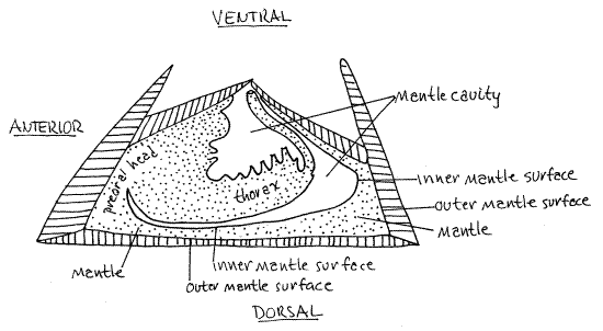
Examine the exterior of the animal. It is completely enclosed by the carapace, or mantle, which is a double fold of the body wall (Fig 1). Barnacles, of course, are arthropods and as such secrete an exoskeleton to enclose the body. In acorn barnacles the outer surface of the double layered mantle is calcified to form a rigid box which contains a soft animal. The remaining exoskeleton, including the inner surface of the mantle, is not sclerotized or calcified and is flexible and thin.
Figure 1. A simplified sagittal section of an acorn barnacle showing the spatial relationships between the body, mantle, and mantle cavity. The animal is upside down with its feet in the “air” and its head stuck to the ground. Barnacle30L.gif
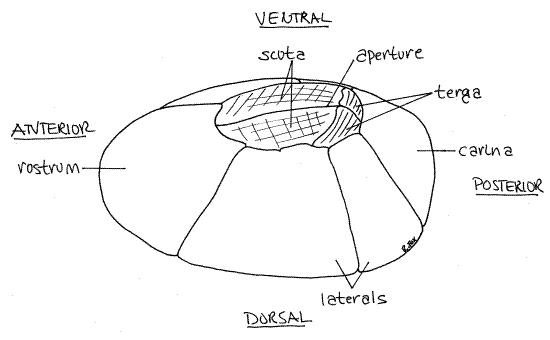
All you can see at present is the heavily calcified part of the exoskeleton. It is underlain by the epidermis of the outer layer of mantle, which secreted it. It is composed of a series of more or less vertically-oriented, overlapping, immovable, calcareous mural plates (= wall plates), which form a ring around the animal, and four movable opercular plates (Fig 2, 19-78B,C).
Primitive barnacles had eight wall plates but modern barnacles have six. The wall plates slope inward and together form a truncated cone resembling a volcano. An opening, theaperture, is at the apex of the cone but it is probably covered by the four opercular plates which form a door, or operculum, to close the aperture. These plates are movable and are operated by specialized muscles.
Balanus has an additional calcareous plate, the basis, which forms the base of the cone and is glued firmly to the substratum (Fig 4, 19-78A). The basis is the floor of the calcareous box inhabited by the barnacle. (Not all acorn barnacles have a calcareous basis. Sometimes it is membranous.) If your specimen has been removed from the substratum, you may be able to see the basis. Sometimes the basis remains with the substratum when the barnacle is removed. Often it remains with the barnacle but is cracked or otherwise damaged. If your barnacle is still attached to the substratum, you will not see the basis at present.
Figure 2. The exterior of a generalized balanomorph barnacle. Barnacle18L.gif
Orientation
This is a good time to orient the animal and determine the major body axes. Acorn barnacles lie on their backs with the dorsal surface attached to the substratum (Fig 1). The base then is dorsal. Look at the aperture and find the four plates forming the operculum. The aperture and operculum are ventral. A long straight cleft, the aperture, divides the operculum into right and left halves, each half being composed of two plates. This line corresponds to the antero-posterior axis, which is the axis of symmetry. Two of the opercular plates are easier to see because their largely horizontal orientation results in a broad exposed surface. These are the scuta, or scutal plates (Fig 2). The scuta are at the anterior end and there is one on the right and one on the left. The other two opercular plates are the terga, or tergal plates. They are posterior. Their orientation is more vertical and consequently you see less of them than of the scuta. Determine right and left.
Mural Plates
The calcareous plates forming the immovable wall of the shell are collectively known as the mural plates (= wall plates). The wall consists of two plates on each side and a single plate at each end. The plates on either side are the lateral plates (Fig 2, 19-78B). Note how they overlap each other. The single plate at the anterior end is the rostrum. It is adjacent to the scuta, of course, and in the genus Balanus, it overlaps the two lateral plates adjacent to it. The carina is the unpaired plate at the posterior end. It is adjacent to the terga and, unlike the rostrum, is overlapped by its adjacent lateral plates (this is not true of all barnacles). The calcareous plates grow with the barnacle and are not shed at each molt although the flexible chitinous portions of the exoskeleton are molted.
Mantle and Mantle Cavity
Use your forceps and/or teasing needles to push the two scutal plates apart and open the aperture of a relaxed barnacle. With low magnification of the dissecting microscope, look into the aperture. The space within the aperture is the mantle cavity. The tough, darkly pigmented lining of the mantle cavity is the carapace, known in barnacles as the mantle. (Even though we have long known that barnacles are not molluscs, this terminology remains appropriate since, in both groups, the mantle is a double layer of body wall, secretes a shell, and encloses a mantle cavity filled with water.)
The mantle is derived from the bivalved carapace of the cypris larva (Fig 19-79B). It is a double fold of the body wall and its inner surface is covered with a thin chitinous exoskeleton (Fig 1). This part of the exoskeleton is not calcified but the outer surface is.
While you are looking into the aperture you will see the thoracopods, or cirri, in their retracted position. These are the thoracic appendages and they are used for suspension feeding. In this position they look like coiled, setose whips. They will be studied in more detail later. Find the cylindrical, transverse scutal adductor muscle running from the inside surface of one scutum across the midline to the other scutum. Its action is to pull the two scuta tightly together and close the aperture. The terga are attached by connective tissue to the scuta so contractions of the adductors move them also and the two sets of plates close in unison.
" Remove the barnacle from the dish and place it on a towel. Use a small (2") C-clamp to rack the articulations between adjacent mural plates. Do this carefully and gently. Place the jaws of the clamp on opposite sides of the shell and apply gentle pressure until you feel the plates give slightly then back off the clamp. Do not apply pressure after the valves move. Inspect the shell and determine which joints are still firm and reposition the clamp to apply pressure to a still solid joint. Continue this until all the plates are loose (The joint between the two left lateral plates need not be broken).
Now place the barnacle in a small dissecting pan of isotonic magnesium chloride (or water, if preserved) and place it on the stage of the dissecting microscope at low power. Use scalpel, needles, fine scissors, and forceps, as required, to remove the rostrum, carina, and right lateral plates from the outer surface of the mantle. Carefully separate the plates from the underlying soft tissue first using scissors to cut the tough mantle and then the scalpel to separate the soft tissue from the plates. Do not cut into the body of the animal.
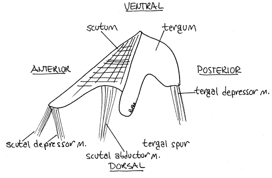
Note that the mantle joins the plates about halfway up their walls. Keep the animal's orientation in mind as you work (Fig 1). Keep the two left lateral plates and the opercular plates intact. They will help support the soft body of the barnacle and also serve as useful landmarks. Look at the left side of the opercular plates. Note the large tergal spur, a process on the lateral border of the tergum. It also makes a useful landmark (Fig 3).
Arrange the animal in the pan so that the exposed right side is up and facing you and the animal's anterior end is to your left. Look at the soft tissue you have exposed. Organs will be studied in the order they appear in the dissection rather than in a standard order or by organ system.
Body Wall
The mantle should still be intact after its calcareous plates have been removed. It is a thin, darkly pigmented in places, layer consisting mostly of epithelium and a thin chitinized cuticle. The soft tissue adjacent to the basis is derived from the greatly enlarged preoral, the remainder of the animal is postoral.
Figure 3. The opercular plates from the left side of an ivory barnacle, Balanus eburneus, from Ft. Pierce, Florida. Barnacle20L.gif
Ovary
The ovary is a large, highly branched, yellowish (in life) organ occupying most of the preoral head along the dorso-lateral length of the animal near the junction of the lateral plates and the basis (Fig 4, 19-78A). It consists of numerous narrow lobes. The other ovary is on the left side but you will not see it now.
>1a. Make a wetmount of a small piece of ovary and use the compound microscope to look for eggs. They should be abundant and yolky. <
Opercular Muscles
Several sets of opercular muscles can be seen at present. Except for the scutal adductor, they originate on the immovable plates of the wall and insert on the movable plates of the operculum. The muscles are white and fibrous but may be covered with a darker connective tissue.
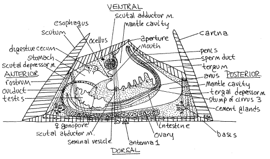
Two scutal depressor muscles originate on the anterior basis and dorsal rostrum to insert on the antero-dorsal corner of the scutum (Fig 4, 19-78A). The scutal abductor muscleinserts on the posterior lateral edge of the scutum.
A powerful tergal depressor muscle extends from the dorsal posterior edge of the tergum to the posterior basis and dorsal carina. These muscles all work together to move the right scutum and tergum laterally and dorsally, thus opening the aperture. An identical set of muscles is present on the left side. These muscles oppose the unpaired scutal adductor muscle, which you have already seen and whose action is to move the right and left scutum and tergum medially and ventrally to close the aperture.
Figure 4. View of the right side of a dissected Balanus eburneus. Barnacle19La.gif
" Use fine scissors to make a longitudinal cut through the right lateral mantle just ventral to the ovary (remember that ventral is toward the aperture, dorsal toward the basis). Extend this cut from anterior to posterior ends of the barnacle. Cut only through the translucent mantle. This will open the mantle cavity from the side.
Reproduction and Development
The mantle cavity may be filled with eggs or shelled nauplius larvae enclosed in a pair of membranous ovisacs secreted by the female system. Eggs are released into the mantle cavity by the female system where they are fertilized by sperm from a nearby individual. The eggs are brooded and naupliar stages are passed in ovisacs in the mantle cavity. Each nauplius is enclosed in a transparent eggshell from which it will eventually emerge before leaving the mantle cavity. Some barnacles retain the larvae in the mantle cavity to the next larval stage, which is the cypris.
Nauplius
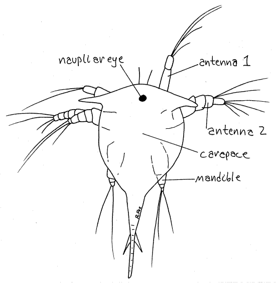
The nauplius larva is the characteristic crustacean larva (Fig 5, 19-79A). It has a large unsegmented head with three pairs of appendages, viz. antenna 1, antenna 2, and mandibles, and a large median naupliar eye. The barnacle nauplius is easily distinguished from other crustacean nauplii by a pair of secretory frontal horns on the anterolateral corners of the head.
>1b. If nauplii are available, either from the mantle cavity of your specimen or from a plankton tow, make a wholemount and examine one with the compound microscope. Find the two pairs of antennae, mandibles, and the naupliar eye. If your nauplius of a barnacle it will have frontal horns. Nauplii removed from the mantle cavity will be enclosed in the eggshell and the horns will not be visible. A few nauplii may have broken from their shells and will be easier to examine. <
Figure 5. The nauplius of an unidentified barnacle species from a plankton tow made at Beaufort, North Carolina. Barnacle21L.gif
Cypris
The cypris is the characteristic barnacle larva (Fig 19-79B). It has a bivalve carapace resembling that of a clam, or more appropriately, an ostracod (Fig 19-85). As in other crustaceans, the carapace is an expanded fold of the body wall of the posterior edge of the head. It is bivalves and its large valves gape ventrally as do those of a clam, ostracod, or cladoceran. The aperture of the adult barnacle develops from the ventral gape of the cypris. Within the two valves is a small crustacean with two compound eyes, a naupliar eye, the first antennae, six pairs of thoracic appendages, and a vestigial abdomen.
The cypris is a type of zoea larva. (Zoea are larvae that swim using thoracic appendages whereas nauplii use head appendages. The zoea stage follows the nauplius, of course.) The lecithotrophic cypris does not feed. It swims for a week or two in the plankton and then settles on a suitable hard substratum. It moves over the substratum using its first antennae as legs. When it finds an acceptable site it attaches using these same antennae and then secretes an adhesive, again using the first antennae, to attach itself permanently. It then undergoes metamorphosis to a miniature barnacle with the shape of the adult. Recently settled barnacles are known as spat.
>1c. If living or preserved cypris larvae are available, examine them and find the structures described above. <
Remove all the reproductive stages and the ovisac from the mantle cavity so you can clearly see the structures within. Remember that the mantle cavity is a small pocket of seawater partly enclosed by the animal but continuous with the sea through the aperture (Fig 1). Don't let the fact that you exposed it by cutting through the body wall mislead you into thinking it is an internal cavity. Demonstrate the continuity between mantle cavity and exterior by passing a needle or probe through the aperture into the space exposed by your lateral incision.
Thorax
It is easiest to study the thorax first and then move anteriorly to the head. Find the large bulbous thorax extending dorsally and posteriorly in the mantle cavity (Fig 4, 19-78A). There is no abdomen.
Direct your attention to the conspicuous cuticularized (but not calcified) biramous thoracic appendages (= thoracopods) arising along the ventral surface of the thorax. Six pairs are present. The appendages extend ventrally into the mantle cavity and, as you know, can be extended from the aperture to feed (Fig 19-80B).
The six pairs of thoracopods are known as cirri and are numbered anterior to posterior. The two similar rami of each thoracopod are long, multiarticulate, and whiplike. The rami bear numerous, long, evenly spaced setae along one edge. The six pairs of cirri have a total of 24 rami. The posterior cirri are the longest and the anterior ones are shorter but more robust. Collectively the cirri form a cast net that is extended from the aperture and swept anteriorly through the water to collect phytoplankton or small zooplankton.
Follow the series of cirri anteriorly from the sixth to the first pair noting that they decrease in length anteriorly but become stouter. The rami of the first pair are the shortest of all.
>1d. Remove a ramus from one of the posterior cirri and make a wetmount of it. Examine it with 100X of the compound microscope and confirm that it is composed of many shortarticles. Each article bears a fringe of long, evenly spaced setae on one edge. Do you think this structure, sweeping through the water in concert with 23 others like it, would make a good filter? <
>1e. Observe a living, unanesthetized barnacle in a dish or aquarium. It may exhibit the characteristic feeding behavior of barnacles. With the aperture open the barnacle extends its setose thoracic appendages in the characteristic "casting" motion barnacles use for feeding. The six pairs of cirri are swept, posterior to anterior, through the water to remove particulate food.<
>1f. Balanus has a pair of light sensitive ocelli and usually responds to a shadow by withdrawing the cirri and closing the aperture. Pass your hand over the living, unaesthetized, feeding barnacles in an aquarium or dish to block the light. <
The anus lies at the bottom of a deep pocket on the dorsal midline just dorsal to the bases of the sixth cirri. The penis also arises from the body wall ventrally between the bases of the sixth cirri. There is no mistaking the penis. It is a long flexible tube capable of achieving impressive lengths when extended. It must be able to reach to other barnacles and deposit sperm in the mantle cavity. An 8-mm barnacle can extend its penis 50 mm to visit a neighbor.
Head
The head and its appendages are anterior to the first pair of cirri of the thorax (Fig 4, 19-78A). It is divided into a large preoral region and a much smaller postoral region. The first antennae are in the preoral region of the head attached to the substratum and the second antennae are absent. Consequently, barnacles have only four pairs of head appendages, three of them mouthparts.
Figure 6. Mouth cone and mouthparts of the goose barnacle, Pollicipes. barnacle16L.gif.
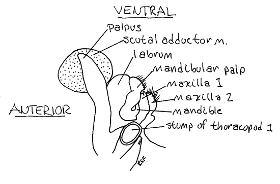
The mouthparts form a mouth cone surrounding the mouth on the ventral midline. Posteriormost among the head appendages are the second maxillae (Fig 6). These are closest to the first cirri and are the posterior border of the mouth cone. They are large and lie closer to the midline than do any other head appendages. Their bases are fused together across the midline. Distally, each bears a dense fringe of long setae on its anterior border.
The first maxillae are smaller than the second and are anterior to and a little lateral to them (Fig 6). Each has a row of short, stout spines on its distal end.
The mandibles are anterior to the first maxillae (Fig 6). Each consists of a base from which arises a large, setose mandibular palp and a smaller medial endite. The endite is tucked out of sight between the palp and the first maxilla. It bears coarse teeth on its distal edge.
The mouth lies on the midline between the two mandibular endites. It is surrounded by the mouthparts and labrum.
A cuticularized bilobed fold of the body wall, the labrum, or upper lip, forms a semicircle around the anterior edge of the mouth (Fig 6). It is not a segmental appendage.
The first antennae are the attachment organs of barnacles and are located on the dorsal surface in the basis, far removed from the other head appendages. You will not see them.
The scutal adductor muscle, which you saw earlier, lies anterior to the head (Fig 4, 6, 19-78A). A tiny eye, which you probably will not see, is located near this muscle. You may have noticed earlier that barnacles are aware of light and dark when you watched it close the aperture and ceased casting when a shadow passed over it.
Each of the two ovaries connects to the exterior by a long oviduct, which is difficult to find. Each oviduct opens into the mantle cavity via a female gonopore located laterally at the base of the first cirri.
The oviducal gland (= egg sac gland) is a swollen, secretory, glandular expansion of the distal end of the oviduct just proximal to the gonopore. It is manifest externally by a large swelling at the base of the first cirrus. The oviducal gland secretes the thin, elastic ovisac which encloses the eggs and embryos in the mantle cavity. The ovisac prevents developing eggs from becoming caught in the feeding current and being swept from the mantle cavity. It gradually deteriorates to allow the escape of the larvae at the appropriate time. The slit-like female gonopore is at the dorsal end of the swelling and is hidden by the overhanging cuticle of the exoskeleton.
Internal Anatomy
Digestive System
The gut is located within the head and thorax and much of it can be seen through the thin body wall (Fig 4, 19-78A). In living material much of the digestive system is pale yellow or brownish. It consists of a mouth, esophagus, stomach, intestine, and anus. You have already seen the mouth and anus. The intestine can be seen through the body wall near the surface on the dorsal, posterior midline of the thorax. It disappears from view beneath the bases of the posterior cirri. The stomach occupies most of the anterior thorax and head and is surrounded by the yellowish digestive ceca, which can be seen without dissection in living material.
" If you wish to expose the gut, proceed as follows. Carefully open the right side of the thorax, using your finest scissors to cut longitudinally through the thin, transparent body wall from posterior to anterior. Do not cut deeper than the body wall.
To find the short esophagus, first remove the head appendages on the right side. Insert the blade of a fine pairs of scissors into the mouth and let it follow the lumen of the esophagus dorsally. When you have the blade in place in the esophagus, close the scissors to cut through the intervening tissue and expose the esophageal lumen. The esophagus is a short, straight tube extending dorsally from the mouth into the anterior end of the stomach.
Anteriorly the stomach is surrounded by the yellowish (in life), lobulated digestive ceca (Fig 4). The stomach occupies much of the preoral head and extends into the anterior thorax.
" Use your fine scissors to extend the existing incision and open the stomach. Cut posteriorly, opening the lumen of the gut as you go. At first you will cut through yellow digestive cecum but posteriorly the stomach is surrounded by the white (in life) testes. Try to avoid cutting the long, curved, bright white (in life) vas deferens.
The stomach is a spacious chamber extending posteriorly to the anterior dorsal corner of the bulge of the thorax (Fig 4). Here it narrows to become the intestine, which runs just beneath the surface, along the posterior edge of the thorax to the anus.
Reproductive System
Most barnacles, including Balanus, are hermaphroditic. You have already seen the penis, ovaries, female gonopores, and maybe the testes. The paired testes are whitish, highly branched tubes draining innumerable tiny spherical testicular follicles where sperm are produced. The testes occupy the anterior thorax and fill most of the space around the gut in this region. They can be seen faintly through the body wall.
Each testis drains via a vas deferens into a large, bright white (in life) seminal vesicle which leads to the base of the penis (Fig 4). It can be seen through the body wall of living specimens.
Each seminal vesicle originates on the ventral surface of the stomach as the fine tubes from the testicular follicles coalesce. It extends posteriorly on the surface of the stomach, then extends along the bases of the cirri to the base of the penis. The right and left seminal vesicles coalesce at the base of the penis to become the sperm duct. This duct extends through the penis to open via the male gonopore at the tip of the penis.
Unlike most arthropods, barnacles have flagellated sperm, a trait that is taken as evidence of their primitive condition.
>1g. Remove some of the testis and vas deferens from the thorax and make a wetmount using seawater. Examine the preparation with 400X and look for sperm. Their long flagella are easily seen at the end of a narrow, elongate head. <
Gill
Return to the vicinity of the left tergal spur and note a large, conspicuous, membranous gill on the mantle. It is composed of large membranous sacs attached to a long central axis. It extends out into the mantle cavity. There is one on the right also and you may have noticed it earlier and wondered what it was. The entire mantle epithelium is thought to function in gas exchange but the gill is specialized for that purpose and has thinner walls than the rest of the mantle. It is hollow and blood circulates through it.
References
Kaestner A. 1970. Invertebrate Zoology, vol. III, Crustacea. Wiley, New York. 523p.
Lochhead JH. 1950. Lepas anatifera, pp 413-418 in Brown FA (ed), Selected Invertebrate Types. Wiley, New York. 597p.
Ruppert EE, Fox RS, Barnes RB. 2004. Invertebrate Zoology, A functional evolutionary approach, 7 th ed. Brooks Cole Thomson, Belmont CA. 963 pp.
Walker G. 1992. Cirripedia. in Harrison, F. W. & A. G. Humes (eds.). Microscopic Anatomy of Invertebrates vol. 9 Crustacea. Wiley-Liss, New York. pp249-312.
Zullo VA. 1979. Marine flora and fauna of the northeastern United States. Arthropods: Cirripedia. NOAA Tech Rep. NMFS Circ 425: 1-28.
Supplies
Dissecting microscope
Compound microscope
Slides and coverslips
Large living (anesthetized) or preserved acorn barnacle
2-inch C-clamp
Isotonic magnesium chloride
Small dissecting pan (sardine tin with wax bottom
Toothbrush
Living specimens should be placed in isotonic magnesium chloride about 60 minutes before needed.
If possible living, active specimens should be available in a dish or aquarium for behavioral observations.