Invertebrate Anatomy OnLine
Sipunculus nudus ©
Peanut worm
4jul2006
Copyright 2003 by
Richard Fox
Lander University
Preface
This is one of many exercises available from Invertebrate Anatomy OnLine , an Internet laboratory manual for courses in Invertebrate Zoology. Additional exercises, a glossary, and chapters on supplies and laboratory techniques are also available at this site. Terminology and phylogeny used in these exercises correspond to usage in the Invertebrate Zoology textbook by Ruppert, Fox, and Barnes (2004). Hyphenated figure callouts refer to figures in the textbook. Callouts that are not hyphenated refer to figures embedded in the exercise. The glossary includes terms from this textbook as well as the laboratory exercises.
Systematics
SipunculaP, Sipunculidea C, Sipunculidae F (Fig 14-14)
Sipuncula P
Sipuncula is a small taxon of 150 described species of marine benthic animals known as peanut worms or star worms. Most are infaunal and wedge themselves into crevices, burrow into soft sediments, or bore into calcareous substrata. Sipunculans are relatively large worms, most being about 10 cm in length but with a range extending from 2 mm to 70 cm.
The anterior end of the body is an eversible introvert with the mouth at its tip. A ring of hollow, ciliated tentacles surrounds the mouth and is used for feeding and gas exchange. The coelom is divided into anterior and posterior compartments. There is no hemal system and the coelom is the fluid transport system. The respiratory pigment, hemerythrin, is present in coelomocytes. The gut is coiled on itself and J-shaped, with a dorsal anterior anus. Two large metanephridia are present. The sexes are separate with gametes maturing in the coelom for later release to the sea through the metanephridia. Cleavage is spiral with a molluscan cross. Development may be direct or with either a trochophore or pelagosphera larva.
Sipunculidea C
The tentacles of sipunculideans form a circle around the mouth whereas those of the sister taxon, Phascolosomatidea C, are dorsal to the mouth. Most are burrowers but some inhabit empty snail shells.
Laboratory Specimens
This exercise is written for Sipunculus nudus, which occurs from North Carolina to Florida on the North American east coast and in California on the west coast. It is present in the West Indies and Bermuda also. Sipunculus is a large worm and individuals may reach 30 cm in length. Sipunculus burrows in muddy sand in shallow coastal waters. Although biological supply houses do not guarantee the specific identity of specimens they ship, Sipunculus is one that is often provided. Phascolopsis gouldi, which is supplied alive by the Woods Hole Marine Biological laboratory, is the subject of another chapter in this series. This exercise is illustrated with drawings of Phascolopsis.
Relaxed living or preserved specimens can be used and, because of the morphological uniformity of the sipunculans, other species can easily be substituted if necessary. If you think you have another species it would be better to use the Phascolopsis exercise as your guide.
External Anatomy
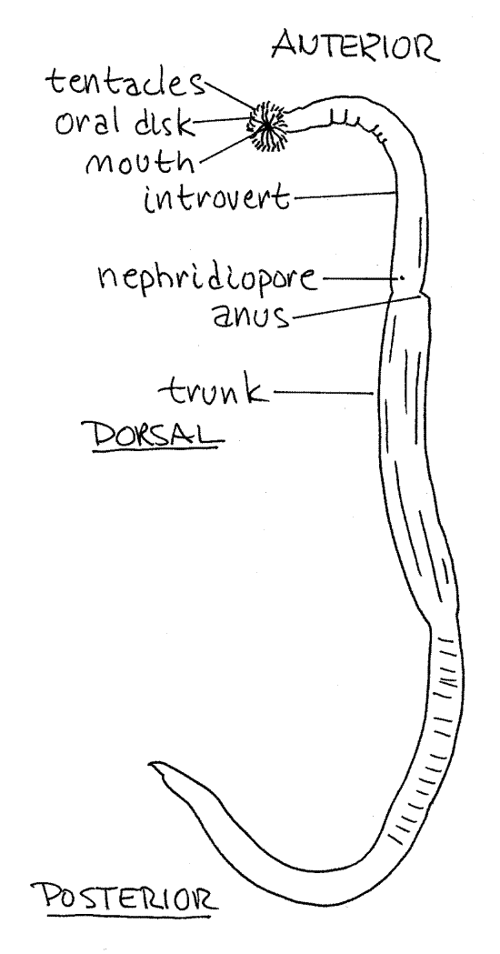
Place a specimen in a long narrow dissecting pan suitable for use with a dissecting microscope and cover it with tapwater. Sipunculus is thick, stout, cylindrical, and wormlike (Fig 1, 14-8A). The body is divided into a short, anterior, retractable introvert and a large posterior trunk (Fig 14-8C) . The introvert is about 1/6 the length of the body and is much smaller in diameter, as well as much shorter, than the trunk. It is covered by small triangular papillae. The introvert can be completely retracted into the anterior trunk by turning it outside in.
Find the anus on the mid-dorsal line between the trunk and introvert. It is a small opening on a low protuberance.
You now have landmarks for anterior and dorsal. Use them to determine posterior, ventral, right, and left. Find the plane of symmetry.
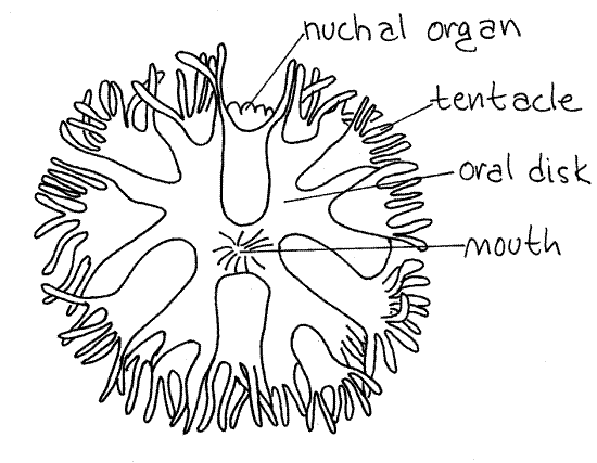
The extreme anterior end of the introvert is the flattened oral disc with the mouth at its center (Fig 2). The mouth and oral disc are surrounded by a ring of small tentacles used for feeding and gas exchange. The number and shape of the tentacles varies with species. The oral disc, mouth, and tentacles cannot be seen if the introvert is even slightly retracted. If the oral disc is not exposed, it will not be visible until the body cavity has been opened.
The surface of the worm is covered by a thick cuticle . The intersections of circular and body wall muscles below the cuticle give Sipunculus a distinctive gridwork appearance.
Two small nephridiopores are located just anterior and ventrolateral to the anus. Sometimes the nephridiopores are on small papillae and easy to find but often the papillae are flattened and the openings are not detectable externally.
Figure 1. The peanut worm, Phascolopsis gouldi, intact and viewed from the left side. Redrawn from Stephen and Edmunds (1972). Sipunc23L.gif
Internal Anatomy
" With fine scissors, make an opening about 3 mm long at the posterior tip of the body to open the trunk coelom. Coelomic fluid will flow from this opening. In preserved specimens the fluid will be clotted
>1a. Place a drop of coelomic fluid on a slide. Use the compound microscope to search for cells. By far the most common are small, transparent, flat, nucleated, spherical hemerythrocytes which contain the pink (in life) respiratory pigment, hemerythrin. The coelomic fluid also contains amoebocytes of various sizes.
Sipunculan gametes mature in the trunk coelom and you may see some. Developing ova are conspicuous, heavy-walled spheres of various sizes. The large nucleus is sometimes visible in the center of these cells. If ova are present, the coelomic fluid will appear distinctly grainy, even to the unaided eye. Sperm are tiny, thimble-shaped cells, each with a long flagellum. Coelomic spermatozoa are typically immobile with inactive flagella, even in living specimens. They are activated by contact with seawater.
Figure 2. En face view of the oral disc of Phascolopsis (after Stephen & Edmunds, 1972 from Andrews, 1890). Sipunc24L.gif
" Place the worm in the center of the dissecting pan with the ventral side down, against the wax of the pan. The anus, which is dorsal, should be facing up, toward you. Insert one point of your finest scissors in the opening made earlier and cut along the dorsal midline anteriorly to the anterior end of the introvert to open the body cavity.
Make this and subsequent incisions using magnification and be careful that you make the incision on the side opposite the nerve cord. Do not cut any organs in the coelom. Try to keep the incision on the dorsal midline using the white nerve cord (Fig 3), which is on the ventral midline, as a landmark. At the base of the introvert, swerve a little to one side to avoid cutting the anus. Pin the animal to the wax of the dissecting pan as you cut, being sure to plan ahead so there will be room in the pan for the extended introvert. Continue the cut all the way to the anterior end of the worm.
" If the introvert is still retracted, and hence outside in, open it anyway. Do this by inserting the tip of your fine scissors in its open (anterior) end and cut posteriorly to the concealed oral disc, stopping when you reach the disc.
The gut and nephridia may have become entangled with each other or with the retractor muscles. If so, attempt to extricate them so they hang freely in the coelom.
At this time you may wish to replace the cloudy water in your dissecting pan with fresh tapwater.
Body Wall
The space opened above is the posterior coelomic compartment, or trunk coelom (Fig 14-10A. The body wall as revealed by your incision consists of a thick external cuticle, thin epidermis, thin connective tissue layer, outer circular and inner longitudinal smooth muscles, and the peritoneum. The epidermal and connective tissue layers are thin, inconspicuous, and are not apparent in gross dissection. The epidermis is a monolayered epithelium that secretes the cuticle and is ciliated only on the tentacles and oral disc. It contains numerous multicellular, secretory epidermal glands which appear as clear microscopic ovals in the cuticle. They are easily seen on the inside surface of the wall of the introvert.
The longitudinal muscle layer of Sipunculus is very thick in the trunk where it is divided by deep grooves into 28-33 longitudinal bundles (Fig 3). The circular muscles are also in bundles. The musculature of the introvert is thinner and is not divided into bundles. Examine the longitudinal muscles with high power of the dissecting microscope. The bundles and the grooves between them are overlain by the thin peritoneum, which is a monolayered, ciliated, squamous epithelium. The circular muscles can be seen just inside the cuticle.
Coelom
The small anterior tentacular coelom is largely confined to the oral disc and tentacles whereas the posterior trunk coelom is much larger and is the major body cavity. The two coeloms are separated by a complete septum in the oral disc. Both are lined by a peritoneum on which cilia occur in tufts on isolated multiciliated cells and both contain hemerythrocytes.
The tentacular coelom is tiny and difficult to see in gross dissection. The central feature of the tentacular coelom is a ring around the pharynx at the base of the oral disc. The ring may, at best, be faintly visible through the body wall at the base of the tentacles. Ciliated tentacular canals from the ring extend into each tentacle and are responsible for the faint pink color of the tentacles of living animals.
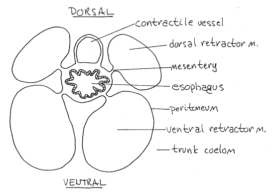
The most obvious feature of the tentacular coelom are the two contractile vessels (Figs 3, 4, 14-10A,B). These are long, pale diverticula extending posteriorly from the tentacular ring into the trunk coelom. They lie on and are attached to the surface of the esophagus in the introvert (Fig 4). In preserved specimens they are white or transparent but may sometimes may contain coagulated hemerythrocytes.
The wall of the contractile vessels are muscular and the peritoneum is ciliated. The muscles cause pulsations of the sac and the cilia generate a bidirectional current to move hemerythrocytes.
The trunk coelom is very large and extends uninterrupted from the oral disc throughout the introvert and trunk. It is not partitioned by septa or mesenteries although small mesenteries are present. The contractile vessels extend posteriorly into the trunk coelom.
Introvert Retractor Muscles
Two pairs of short introvert retractor muscles extend from the body wall of the trunk to the anterior end of the introvert. Their contractions pull the introvert into the trunk. It is re-extended by the action of the body wall muscles on the coelomic fluid.
Figure 3. Dorsal dissection of Phascolopsis gouldi. Sipunc25L.gif
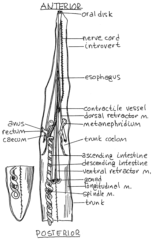
The four muscles are about the same length in Sipunculus. They arise on the body wall at about the level of the anus and extend anteriorly to the oral disc. The dorsal introvert retractor muscles originate on the dorsal body wall whereas the ventral introvert retractor muscles arise on the ventral wall (Fig 2, 14-10A).
The retractor muscles are long, flat, white, strap-like muscles derived from the longitudinal muscles of the body wall. Use a higher power to examine the posterior end of a dorsal retractor muscle and confirm that it is a continuation of the longitudinal body wall muscles.
Anteriorly the four retractor muscles are connected to each other by connective tissue and form a sheath around the anterior gut and contractile vessel (Fig 4).
Figure 4. Cross section of the introvert of Phascolopsis (redrawn from Cuénot, 1900). Sipunc26L.gif
Digestive System
The J-shaped sipunculan gut forms a long coiled loop between the mouth and anus. The two arms of the loop are twisted tightly around each other.
The mouth is in the center of the oral disc. It opens into a short, wide pharynx in the anterior introvert. Posteriorly the pharynx constricts to become the long, narrow, straight esophagus that extends the length of the introvert to the anterior trunk. The dark contractile vessels lie on the surface of the pharynx and esophagus. Each retractor muscle is attached to the esophagus by a mesentery (Fig 4).
In the anterior trunk the esophagus becomes the stomach which initiates a short series of loose coils to form the 'sipunculus loop" a characteristic feature of the family Sipunculidae. This loop is not present in Fig 2 but can be seen in 14-10A.
The exceedingly long, helically coiled intestine exits the stomach and extends far posterior in the trunk as the descending intestine. It then reverses direction and climbs back into the anterior trunk as the ascending intestine (Fig 3). In some specimens it may be filled with sand and distended. The descending and ascending intestine are coiled around each other to form the intestinal loop which is characteristic of all sipunculans, not just Sipunculus.
The intestinal loop consists of about 15 double coils around a long, slender, central spindle muscle (Fig 3. 14-10A). This muscle originates on the body wall near the anus, supports the intestine, and shortens the intestinal loop when it contracts. It inserts on the posterior bend of the intestine (not on the body wall) and is attached to the ascending intestine by a mesentery.
The intestinal loop of Sipunculus is attached to the body wall by numerous small mesenteries.
In the anterior trunk cavity the ascending intestine ceases coiling and becomes the short, straightrectum which joins the body wall on the anterior, dorsal midline at the anus (Fig 3). There is a tiny, bulbous cecum at the point where the intestine becomes the rectum. In some specimens the cecum may be very long and extend posteriorly for about half the length of the intestinal loop. It is white in preserved material. The rectum is attached to the body wall by a small mesentery. In Sipunculus a pair of racemose glands of unknown function form tufts beside the rectum.
The faint yellowish color of the intestine of living specimens is due to the presence of a thin layer of chlorogogen cells in the mesothelium. The mesothelium covering the ascending intestine bears numerous ciliated pits called fixed urns but they are not apparent in gross dissection. Chlorogogen cells associated with the urns accumulate foreign particles from the coelomic fluid.
Respiratory and Fluid Transport Systems
The tentacles of the oral disc probably are the chief respiratory surface and the tentacular coelom functions as a fluid transport system for the distribution of oxygen. Hemerythrin in the tentacular coelom is oxygenated in the tentacles and the hemerythrocytes moved by the ciliated peritoneum to the contractile vessel where the oxygen is transferred to hemerythrocytes of the trunk coelom for distribution to the tissues.
In some sand-dwelling sipunculans, Sipunculus included, integumentary coelomic dermal canals extend from the trunk coelom into the integument where they run between muscle bundles near the surface. Coelomic fluid circulates through them and they are thought to function in gas exchange and distribution.
Excretory System
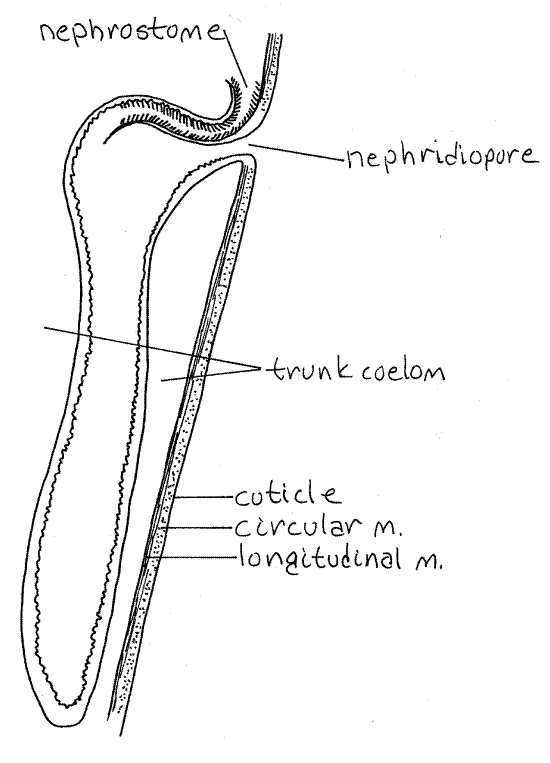
The two large, dark brown metanephridia are long, narrow sacs that hang freely in the coelom (Fig 3). They are attached to the anterior lateral body wall and open to the exterior via the nephridiopores just anterior to the anus. If you could not find the nephridiopores earlier, unpin this region of the worm and find them now. The nephrostome is an opening from the trunk coelom into the nephridium (Fig 5). It is located on the dorsal side of the nephridium at the point where the nephridium attaches to the body wall and is covered by a small, transparent, semicircular lip. Most of the nephridium is a long, contractile, blind sac in which mature gametes are stored. The nephridia function as gonoducts through which the gametes exit the coelom.
Nervous System
A conspicuous, white ventral nerve cord lies exposed on the ventral midline of the trunk coelom (Fig 3). Look for the unpaired lateral nerves that exit it at irregular intervals to enter the body wall. Use high power of the dissecting microscope to look for the lateral nerves. These nerves are evident along the entire length of the nerve cord and can be seen best by slightly lifting the cord with the tiny needle.
There are no ganglionic swellings of the nerve cord and no hint of segmental arrangement of the lateral nerves. The ventral nerve cord extends for the length of the coelom and bifurcates ventral to the anterior pharynx to form two circumenteric connectives that enter the wall of the gut and extend dorsolaterally around the pharynx at the base of the tentacles.
Figure 5. Section through a metanephridium of Phascolopsis. sipunc27L.gif
The two connectives form a nerve ring around the pharynx. They join dorsally at the white bilobed brain which lies on the midline in the roof of the pharynx between the insertions of the two dorsal retractor muscles. Two tiny black eyespots are embedded in the dorsal surface of the brain and will help you find the brain. The brain is partly surrounded by the tentacular coelom which presumably supplies it with oxygen.
The nerve ring and brain are imbedded in connective tissue and are difficult to expose but in living specimens pink corpuscles in the sinus may make the brain region easier to locate. It is recommended that you not attempt to uncover these structures, rather view them as best you can through the surrounding tissues. Both can be seen faintly through the retractor muscle sheath covering the base of the tentacular crown.
Reproductive System
Fertilization is external and, although sipunculans are gonochoric, there is no sexual dimorphism. Fertilization is external. The two gonads are inconspicuous, frilly, transverse ridges across the base of the two ventral retractor muscles (Fig 3). Germ cells do not ripen in the gonads nor are gametes stored here so the gonads are never large.
Developing germ cells are released into the trunk coelom where they complete their maturation and become spermatozoa or ova. Mature gametes accumulate in the metanephridia prior to release. They are then forced out the nephridiopore by contractions of muscles in the nephridial wall. Spiral cleavage produces a trochophore larva.
References
Andrews EA . 1890. Notes on the anatomy of Sipunculus gouldii Portuales. Stud. Biol. Lab. Johns Hopkins Univ. 4:389-430, pls. 44-47.
Brown FA . 1950. Phascolosoma gouldii, pp 309-317 in Brown FA (ed.) Selected Invertebrate Types. Wiley, New York. 597p.
Cuenot L. 1900. Le Phascolosoma commun (Phascolosoma vulgare de Blainv.) [in] Boutan, L. (ed.) Zool. descriptive des invertebres, Paris1:386-422.
Cutler EB. 1973. Sipuncula of the western North Atlantic . Bull. Am. Mus. Nat. Hist. 152(3):105-204.
Cutler EB. 1994. The Sipuncula Their systematics, Biology, and Evolution. Cornell Univ. Press, Ithaca. 453 pp.
Fisher WK. 1950. The sipunculid genus Phascolosoma. Ann. Mag. Nat. Hist. ser 12, 3:547-552.
Hyman LH . 1959. The Invertebrates: Smaller Coelomate Groups, vol. V. McGraw-Hill, New York, 783p.
Kohn AJ, Rice ME. 1971. Biology of Sipuncula and Echiura. Bioscience 21:5833-584.
Metalnikoff S. 1900. Sipunculus nudus. Z. Wiss. Zool. Abt. A 68:261-322.
Peebles F, Fox D. 1933. The structure, functions, and general reactions of Dendrostoma zostericola. Bull. Scripps Inst. Oceanogr. Ser 3:201-224.
Pilger JF. 1982. Ultrastructure of the tentacles of Themiste lageniformis (Sipuncula). Zoomorph. 100:143-156.
Rice ME. 1981. Larvae adrift: Patterns and problems in life histories of sipunculans. Amer. Zool. 21:605-619.
Rice ME, Todororic T , eds. 1970. Proceedings of the International Symposium on the Biology of the Sipuncula and Echiura. Inst. Biol. Res. Yugoslavia and Smithsonian Institution, Washington. Vol. 1 (355p), vol. 2 (254p).
Stephen AC, Edmonds SJ . 1972. The Phyla Sipuncula and Echiura. British Mus. Nat. Hist, London. 528 p.
Ruppert EE, Fox RS, Barnes RB. 2004. Invertebrate Zoology, A functional evolutionary approach, 7 th ed. Brooks Cole Thomson, Belmont CA. 963 pp.
Supplies
Compound microscope
Dissecting microscope
Dissecting pan (kippered herring tin or aluminum ice tray with wax bottom)
Dissecting set with microdissecting tools
Isotonic magnesium chloride if using living specimens
Seawater if using living specimens
Living or preserved Sipunculus
#1 stainless steel insect pins