Invertebrate Anatomy OnLine
Myzostoma ©
Crinoid Commensal
4jul2006
Copyright 2003 by
Richard Fox
Lander University
Preface
This is one of many exercises available from Invertebrate Anatomy OnLine , an Internet laboratory manual for courses in Invertebrate Zoology. Additional exercises, a glossary, and chapters on supplies and laboratory techniques are also available at this site. Terminology and phylogeny used in these exercises correspond to usage in the Invertebrate Zoology textbook by Ruppert, Fox, and Barnes (2004). Hyphenated figure callouts refer to figures in the textbook. Callouts that are not hyphenated refer to figures embedded in the exercise. The glossary includes terms from this textbook as well as the laboratory exercises.
Systematics
AnnelidaP, Polychaeta C, Palpata, Aciculata, Myzostomida O, Myzostomidae F (Fig 13-7A)
Annelida P
Annelida consists of the segmented worms in the major taxa Polychaeta (bristleworms), Oligochaeta (earthworms and relatives), Branchiobdellida (crayfish ectosymbionts), and Hirudinea (leeches) with a total of about 12,000 known species in marine, freshwater, and terrestrial environments. The segmented body is composed of an anterior prostomium, a linear series of similar segments, and a posterior pygidium. The prostomium and pygidium are derived from anterior and posterior ends of the larva whereas the intervening segments arise through mitotic activity of mesodermal cells in the pygidium.
The body wall consists of a collagenous cuticle secreted by the monolayered epidermis. A connective tissue dermis lies beneath the epidermis. The coelom is lined by a mesothelium which may be specialized to form the body wall muscles. Most annelids have chitinous bristles, or chaetae, secreted by epidermal cells, that project from the body. The coelom is large, segmentally compartmented, lined by mesothelium, and well developed in polychaetes and oligochaetes but reduced in leeches. Successive coelomic spaces are separated by transverse bulkheads known as septa which consist of double layers of mesothelium with connective tissue in between. The right and left sides of each segmental coelom are separated by longitudinal mesenteries which, like septa, are double layers of mesothelium with connective tissue between.
The gut is a straight, regionally specialized tube that begins at the mouth at the anterior end and extends for the length of the body to end at the anus on the pygidium. It penetrates each septum and is supported by dorsal and ventral mesenteries. Like that of most invertebrates, the gut consists of ectodermal foregut, endodermal midgut, and ectodermal hindgut. The nervous system consists of a dorsal brain in or near the prostomium, a pair of circumpharyngeal connectives around the anterior gut, and a double, ventral nerve cord with paired segmental ganglia and nerves. The hemal system of most annelids is a set of tubular vessels, some of which are contractile and serve as hearts. The hemal system is absent or greatly reduced in leeches. The system includes a dorsal longitudinal vessel above the gut in which blood moves anteriorly, a ventral longitudinal vessel below the gut, in which blood moves posteriorly, and paired segmental vessels that connect the dorsal and ventral vessels. The digestive, hemal, and nervous systems are continuous and pass through the segments.
Respiration is accomplished in a variety of ways. In some, the general body surface is sufficient but gills are present in most polychaetes, many leeches, and a few oligochaetes. Excretory organs are metanephridia or protonephridia and typically one pair is present in each segment. These osmoregulatory organs are best developed in freshwater and terrestrial species. The sexes are separate in polychaetes but oligochaetes and leeches are hermaphroditic. In the ancestral condition paired submesothelial clusters of germ cells were present in each segment and released developing gametes into the coelom. In derived taxa reproductive functions tend to be confined to a few specialized genital segments. Gametes mature in the coelom or its derivatives and fertilization is external. Gametes are shed through ducts derived from metanephridia or by rupture of the body wall. Spiral cleavage follows fertilization. Clonal reproduction is common.
Polychaeta C
Polychaeta is a large (8000 species) and diverse taxon of marine annelids thought to be the most primitive of the annelid taxa and the most like the ancestral annelid. The body of a typical polychaete is divided into segments, each of which bears a pair of fleshy appendages, or parapodia. The head is often equipped with abundant, well-developed sense organs. The anterior gut is muscular, sometimes eversible, and frequently equipped with chitinous jaws. Polychaetes are gonochoric and gametes ripen in the coelom from which they are shed through ducts or by rupture of the body wall.
Palpata
The prostomium has a pair of sensory palps. These are lacking in the sister taxon, Scolicida.
Aciculata
Aciculata is a large taxon containing many worms well-known to marine biologists and invertebrate zoology students. The parapodia are well-developed and biramous. The prostomium has antennae. Aciculates were once known as “errant” polychaetes because they are active and mobile.
Myzostomida O
Myzostomes are small (5 mm) derived annelid worms usually considered to polychaetes, although their sister taxon is unknown and they are sometimes placed in Gnathifera. All are parasites of echinoderms, primarily crinoids. They remove food from the food grooves of their hosts. The 100 species belong to the genus Myzostoma in Myzostomidae. The body consists of five segments with five pairs of parapodia. The coelom is unsegmented and no hemal system is present. Myzostomes are hermaphroditic, usually consecutive. Ciliated coelomic channels extend from the coelom to the gut but they are not thought to be excretory.
Laboratory Specimens
Myzostoma is rarely studied, either alive or preserved, in introductory invertebrate zoology courses. It may occasionally be available at coastal laboratories and biological stations in areas where feather stars are found.
External Anatomy
Examine a living or preserved specimen of any species of Myzostoma. Living specimens should be observed in seawater and then placed in isotonic magnesium chloride (see Techniques chapter). The body is strongly flattened dorsoventrally and is oval or round in dorsal view (Fig 1, 13-42A). They exhibit few signs of segmentation internally or externally. The dorsal surface is essentially featureless although it may be ridged or furrowed. Ten pairs of narrow short sensory cirri, perhaps homologous to the cirri of the parapodia of other polychaetes, extend from the margins of the disk-like body.
Make a wetmount of a specimen with its ventral surface uppermost. Support the coverslip with wax feet to protect the worm (See Techniques chapter). Apply a little pressure to the wax so the animal is squeezed slightly.
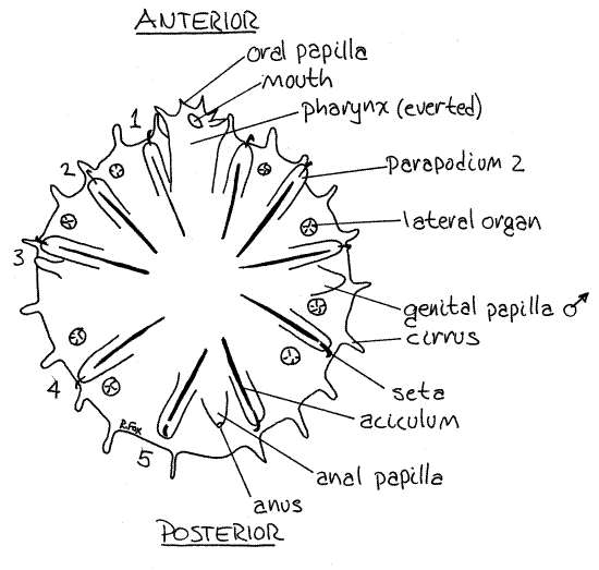
Observe the ventral surface with the compound microscope. It has five pairs of uniramous parapodia. These are short fleshy cylinders, each bearing a stout hooked chaeta at its distal end. The appendage is stiffened by a long chitinous aciculum extending from the core of the parapodium into the body.
The gut extends from the anterior mouth to the posterior anus (Fig 13-42A). Extensive diverticula, the digestive ceca, extend from the gut throughout the body. These may be visible in some specimens. The anterior end of the gut is a muscular pharynx which can be everted through a small pore in the body (Fig 1). When everted, as it often is, it can be seen to have a ring of papillae around the mouth at its distal end (Fig 1). The anus is on a small anal papilla at the posterior end of the worm (Fig 1). The gut runs straight from mouth to anus.
Four pairs of sucker-like lateral organs that are thought to be sense organs are located laterally on the ventral surface of the body beside the parapodia.
The male gonopores are located on a pair of genital papillae, one at the base of each third parapodium. The extensive testes are scattered throughout the coelom. The ovaries empty via an oviduct into the posterior gut. The ovaries are also associated with the coelom.
Figure 1. Ventral view of a Myzostoma from on the feather star Comactinia echinoptera collected at St. Lucie Rocks, Florida. The pharynx is everted. The segments are numbered. Poly84L.gif

References
Hyman LH. 1955. The Invertebrates: Echinodermata, IV. McGraw-Hill, New York. 763pp.
Jägersten G. 1939a. Zur Kenntnis der Larvenentwicklung bei Myzostomum. Ark. Zool. 31A.
Jägersten G. 1939b. Uber die Morphologie und Physiologie des Geschlechtsapparatus und den Kopulationmechnaismus der Myzostomiden. Zool. Bidr. fran Uppsala, 18.
Jägersten G. 1940. Zur Kenntnis der Morphologie, Entwicklung und Taxonomie der Myzostomida. Nova Acta Regiae Soc. Sci. Upsalla, ser 4, 11(8)1-84.
Kozloff EN. 1990. Invertebrates. Saunders College Pub., Philadelphia. 866p.
Pettibone MH. 1982. Annelida, in Parker SP. (ed). Synopsis and Classification of Living Organisms, vol. 2. McGraw-Hill, New York. pp. 41-42.
Ruppert EE, Fox RS, Barnes RB. 2004. Invertebrate Zoology, A functional evolutionary approach, 7 th ed. Brooks Cole Thomson, Belmont CA. 963 pp.
Supplies
Dissecting microscope
Living or preserved Myzostoma
Sea water and isotonic magnesium chloride