Invertebrate Anatomy OnLine
Hydra ©
24may2007
Copyright 2001 by
Richard Fox
Lander University
Preface
This is one of many exercises available from Invertebrate Anatomy OnLine , an Internet laboratory manual for courses in Invertebrate Zoology. Additional exercises can be accessed by clicking on the links to the left. A glossary and chapters on supplies and laboratory techniques are also available. Terminology and phylogeny used in these exercises correspond to usage in the Invertebrate Zoology textbook by Ruppert, Fox, and Barnes (2004). Hyphenated figure callouts refer to figures in the textbook. Callouts that are not hyphenated refer to figures embedded in the exercise. The glossary includes terms from this textbook as well as the laboratory exercises.
Systematics
Cnidaria P, Medusozoa, Hydrozoa C, Anthoathecatae O (Fig 7-75, 7-74)
Cnidaria P
The cnidarian body consists of a central blind sac, the coelenteron (= gastrovascular cavity), enclosed by a body wall comprising two epithelia, the outer epidermis and the inner gastrodermis (Fig 7-1, 7-2*). A gelatinous connective tissue layer, the mesoglea, lies between the two epithelia. The mouth opens at one end of the coelenteron and marks the oral end. The mouth is at the tip of a process, the manubrium that elevates it above the oral surface. The opposite pole is the aboral end. The imaginary line connecting the oral and aboral poles is the axis of symmetry around which the radial symmetry of the body is organized. The mouth is usually surrounded by one or more circles of tentacles.
The defining cnidarian feature is, of course, their possession of stinging cells, or cnidocytes (Fig 7-8). Characteristic of the epidermis, they are also sometimes found in the gastrodermis. Cnidocytes contain an explosive organelle, the cnida, which, upon proper stimulation, inverts and ejects a slender, often barbed and toxic thread in the direction of prey or predator (Fig 7-9). Three types of cnidae are found in cnidarians (Fig 7-10). Nematocysts (in nematocytes), spirocysts (in spirocytes), and ptychocysts (in ptychocytes). All toxic cnidae are nematocysts whereas spirocysts are sticky, and the everted tubules of ptychocysts are used for constructing feltlike tubes. Most cnidae are nematocysts and these are present in all three higher cnidarian taxa. Spirocysts and ptychocysts are found only in Anthozoa.
The basic body plan described above can be manifest as a swimming medusa or attached polyp. In some taxa only one generation is present whereas in others both are found. A life cycle featuring alternation of sexual, swimming medusae with benthic asexual polyps is typical of many cnidarians.
All cnidarians are carnivores feeding on live prey which they usually capture using tentacles armed with cnidocytes. Digestion occurs in the coelenteron which is typically equipped with ciliated canals for distribution of partly digested food. Cnidarians are ammonotelic and diffusion across the body and tentacle surface eliminated the ammonia from the body. Gas exchange is across the general body surface. The nervous system is a plexus of basiepithelial neurons serving sensory and motor systems (Fig 7-6). Most cnidarians are gonochoric. The life cycle typically includes a planula larva. Cnidarians are chiefly marine but the well-known Hydra is an exception.
Medusozoa
Medusozoa comprises those cnidarians whose life cycle includes a medusa generation that alternates with a polyp generation (Fig 7-75B). Symmetry is radial and tetramerous. Nematocysts are the only type of cnidocyte present. Included taxa are Scyphozoa (jellyfishes) and Hydrozoa (hydroids, Hydra, Portuguese men of war, etc).
Hydrozoa C
Hydrozoa is a diverse taxon of about 3000 species of mostly marine cnidarians. The life cycle usually includes both polyp and medusa generations (Fig 7-65A) but may be entirely polyp (Fig 7-65B) or entirely medusa (Fig 7-65C). Polyps typically are colonial and medusae usually solitary. Some form colonies of combinations of polyps and medusae. The few freshwater cnidarians, such as Hydra, Vallentinia, and Craspedacusta, are hydrozoans.
Hydrozoan polyps are usually small, about 1 mm in length, and colonial. Hydromedusae are also small, at least in comparison with scyphomedusae, and are usually less than 1 cm in diameter. Hydromedusae are further distinguished from scyphomedusae by possession of a velum, a circumferential shelf of tissue that encircles the subumbrellar concavity and functions as an adjustable diaphragm to create a pulse of water for swimming. Medusae are tetramerously symmetrical as are scyphomedusae and four radial canals and four tentacles are usually present.
Cnidocytes are found only in the epidermis and germ cells arise in the gastrodermis but may, or may not, migrate to the epidermis to form gonads and mature into gametes. Nematocysts are the only cnida. Gonads may be on the manubrium or on the radial canals. Gametes, even if gastrodermal, are released directly to the surrounding water, never, in contrast with scyphomedusae, into the coelenteron.
Anthoathecatae O
Hydra is an unusual anthoathecate hydrozoan. It is a freshwater species in which the medusoid generation is absent and the polyps solitary. There is no planula and zygotes develop into shelled embryos which leave the female and develop directly into polyps (Fig 7-65B). It is athecate, lacks periderm, and has filiform tentacles although it belongs to a taxon, Capitata, most of whose members have capitate tentacles (with a terminal swelling).
Laboratory Exercises
This exercise utilizes living specimens available inexpensively from biological supply companies and commercially prepared slides, available from the same source. Cross sections, longitudinal sections, and wholemounts, are available as well as sections and wholemounts of specimens with ovaries and testes. Slides can be used to supplement, or if necessary replace, study of living specimens.
Gross Anatomy
Begin the exercise with observations of general anatomy. Place a drop of water containing a single living Hydra in the center of a clean, dry 6-cm culture dish.
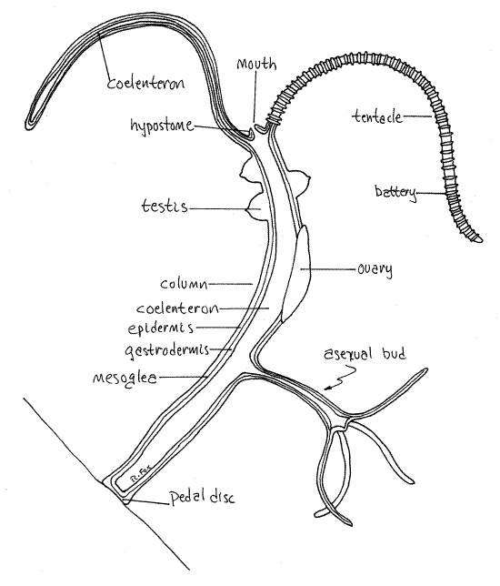
Using the dissecting microscope as necessary, examine the specimen after giving it time to relax and extend its column and tentacles. Hydra consists of an elongate column inside of which is the coelenteron Fig 1, 7-57*). The mouth is located at the oral end atop a low mount, the hypostome. It is surrounded by a ring of hollow tentacles, which are well-endowed with cnidocyte batteries. The animal will probably attach to the glass with its adhesive pedal disc, located at the aboral end. Hydra sometimes secretes a gas bubble from the pedal disc and floats away to find a new substratum.
Most Hydra are gonochoric and both sexual and asexual (clonal) reproduction occur. Asexual reproduction is via asexual buds that form on the parent animal. They look like, and are, small hydras (Fig 1, 7-57) that will separate from the parent and adopt an independent existence. Hydra does not form colonies.
Gonads form in the epidermis of mature animals. These are either ovaries or testes which produce eggs or sperm, respectively, by meiosis. Gonads are low conical mounds on the surface of the column, ovaries are larger than testes. Testes have a central pore from which sperm escape and swim to a waiting ovum in the ovary of a female. Fertilization results in a zygote which develops a chitinous shell and leaves the ovary. Look for asexual buds or gonads on your specimen.
Feeding Behavior
As in most cnidarians, food is captured by the tentacles, stung with the cnidocytes, and then transferred to the mouth and coelenteron for initial extracellular digestion. This is followed by intracellular digestion as molecules and food particles are distributed by gastrodermal ciliary currents in the coelenteron and then endocytosed by gastrodermal cells. Undigested food vacuoles are exocytosed into the coelenteron and then voided through the mouth, which is thus the anus also. Notice again the cnidocyte batteries on the tentacles. The mouth in hydra is temporary, being present only when there is food to ingest. At other times the cells surrounding the mouth position adhere to each and the mouth disappears.
Place 2 or 3 brine shrimp nauplius larvae in the dish and watch the animals with the dissecting microscope. Note the effect of contact with tentacles on the prey. Based on your observations, do you think the nematocysts are toxic or sticky? How long does it take for the prey to succumb? Does Hydra actively stalk its prey or does it seem to wait for food to blunder into its tentacles? How are the brine shrimp transferred to the mouth and coelenteron?
Microscopic Anatomy
Figure 1. Hydra drawn with a combination of testes, ovary, and asexual bud. Cnidocyte batteries are shown on only one tentacle. Hydrozoa79L.gif
Place a living Hydra on a microscope slide with a small drop of water. Place the slide on the stage of the compound microscope, without a coverslip, and observe the animal with 100X. Do not try to use high power without a coverslip. Observe the morphology of the animal relocate the column, pedal disk, tentacles and mouth (if present).
Look closely at the surface of your Hydra to see the conspicuous spherical nematocysts. These are the explosive capsules of cnidocytes. The cnidocytes themselves will probably not be discernable but their capsules are abundant and
conspicuous. There are two sizes, one much smaller and more abundant than the other. Find one or more of the large ones that can be observed in side view and look for the trigger, orcnidocil, protruding from the exposed edge of the capsule.
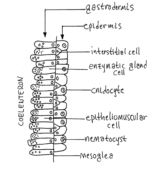
Place a coverslip on your preparation and observe the animal with 400X. The Hydra is now distorted but you will be able to see some things that you could not see before. Look at the edge of the column. A thick monolayer of epidermal cells should be evident (Fig 2). Many of these are cnidocytes containing nematocysts.
Just inside the epidermis you will see a thin dark line, the mesoglea. Remember that the mesoglea of polyps is very thin and acellular. The next layer is the gastrodermis but this will be difficult to discern in wholemounts. There are simply too many cells in the way.
Look at one of the tentacles with 400X. Note the two sizes of nematocysts. Most of the nematocysts will be intact and unexploded but some will have discharged. Find some of the large ones, focus carefully, and look for a coiled thread inside the capsule. If the thread is present, the nematocyst is undischarged. Look around for some discharged nematocysts. These will look quite different. They are obviously empty, having everted their thread, which can be clearly seen extending away from one end of the empty capsule. With careful focusing and light adjustment you can also see the formidable barbs at the base of the thread adjacent to the empty capsule.
Place a drop of 1% acetic acid beside the coverslip and draw it under while watching through the microscope. The acid may stimulate the discharge of many of the nematocysts and, if you are fortunate, you may actually see one of them discharge as you watch.
Prepared Slides
Sections
Anatomy
Use the compound microscope to study stained commercially prepared slides of cross and longitudinal sections of Hydra. Use the scanning lens to search the slide and locate the longitudinal section. Once found, use 100X to begin your study.
Allowing for variations due to the location of the plane of section, you should see a cylinder of stained cells. The cylinder should be closed at both ends. The cylinder is the column (Fig 1).
Tentacles, variously sectioned along transverse, oblique, or longitudinal planes, should also be visible around one end of the column. The end of the column with the tentacles is the oral pole and an opening, the mouth, may be visible. In most specimens the mouth is closed and even if open the plane of section probably does not pass through it.
The open space in the interior of the column is the coelenteron. It may contain food items and parts of it may be occluded by the gastrodermis.
The animal is longitudinally divisible into four regions which should be discernable in your section. At the oral pole is the hypostome, which is the region surrounding the mouth. The tentacles arise in a circle around the base of the hypostome.
Below the hypostome is the stomach region. This region accounts for most of the column. Below it is the shorter, often constricted, stalk. Finally, at the extreme aboral pole, is thepedal disk by which the animal attaches to the substratum.
Histology
Use 400X to study the histology of the body wall (Fig. 2). This wall consists of two epithelia, epidermis and gastrodermis, separated by a thin, acellular, connective tissue layer. The connective tissue is the mesoglea and it should be visible over most of the section as a thin dark line. The mesoglea is little more than the basal laminae of the two epithelia. The two epithelia are monolayered (or pseudostratified) and consists of a layer of cuboidal or columnar cells.
Study the histology of the two epithelia in more detail. Make the study at 400X with the compound microscope. Cell membranes cannot be seen in these preparations but nuclei and other organelles may be visible. You will not be able to distinguish among the cell types present.
Figure 2. A longitudinal section through the body wall of the column of Hydra. Hydrozoa78L.gif
Epidermis
The epidermis covers the outside of the animal and in almost all regions (except the pedal disk) it is the thinner of the two epithelia. It contains epitheliomuscular cells with longitudinal fibers, cnidocytes, sensory cells, and neurons.
The epidermis a relatively thin layer in which you can see spherical or ovoid cell nuclei and nematocysts. The nuclei have one or two dark-staining nucleoli whereas the nematocysts are translucent with a central coiled thread. Nematocysts are most abundant in the hypostome and tentacles.
Although cell membranes are not apparent, you can get an idea of the number of cells present by the abundance of nuclei. There are many more cells than there initially appear to be.
There is a thin secreted layer, the periderm, covering the apical side of the epidermis. In some hydrozoans this is elaborated to form a perisarc.
The epidermis of the pedal disc is a thick, monolayered columnar epithelium consisting largely of secretory cells that produce the adhesive used to hold the animal to the substratum.
Gastrodermis
Inside the mesoglea, lining the coelenteron, is the thicker gastrodermis. It includes epitheliomuscular cells with circular muscle filaments, enzymatic gland cells cells, neurons, and sensory cells. As mentioned earlier, the histology of the four regions of the column differ and these differences are most evident in the gastrodermis. The gastrodermis of the hypostome is a thick pseudostratified or monolayered (simple) columnar epithelium. Some of these are mucus-secreting gland cells and others are epithelionutritive cells for absorption.
The stomach region has a thick gastrodermis that includes many enzymatic gland cells, whose vacuoles contain zymogens (inactive form of enzymes), and nutritive epitheliomuscular cells whose vacuoles contain food particles undergoing intracellular digestion.
The gastrodermis of the stalk region is dominated by abundant nutritive epitheliomuscular cells with very large vacuoles. The gastrodermis of the pedal disk is a thin, monolayered, cuboidal epithelium.
Reproduction
Ovary Wholemount. Use 40X and 100X to study the wholemount of an adult hydra with an ovary (Fig 1). How many ovaries are present? _________
Ovary cross section. Use 100X and 400X to study the cross section of an ovary. Find an ovary and study its histology. It is located in the epidermis, on the outside of the mesogleabut its germ cells originated in the gastrodermis. A multilobed developing ovum is located within the mass of epidermal cells. The lobes are more or less circular in section. Depending on the plane of section you may see the nucleus in one of the lobes.
Testis Wholemount. Use 40X and 100X to study the wholemount of an adult hydra with a testis (Fig 1). How many testes are present? ____. Why might an individual have many testes but only one ovary?
Testis cross section. Use 100X and 400X to study the testis cross section. Find the mesoglea and note that the testis is outside it, thus in the epidermis, although the germ cells originate in the gastrodermis. Find the thin layer of epidermal cells covering the mass of germ cells in various stages of spermatogenesis, including spermatozoa. The testis is partly partitioned by septa.
Asexual bud wholemount. Use 40X and 100X to study the wholemount of an adult hydra with an asexual bud (Fig 1). How many buds are present ____
*Hyphenated call-outs, such as this one, refer to figures in Ruppert, Fox, and Barnes (2004). Those without hyphenation refer to figures embedded in this exercise.
References
Freeman WH, Bracegirdle B . 1971. An atlas of invertebrate structure. Heinemann Educational Books, London.
Hyman LH. 1940. The Invertebrates: Protozoa through Ctenophora, vol. I. McGraw Hill, New York. 726 p. See page 374.
Ruppert EE, Fox RS, Barnes RB. 2004. Invertebrate Zoology, A functional evolutionary approach, 7 th ed. Brooks Cole Thomson, Belmont CA. 963 pp.
Supplies
1 dissecting microscope
1 compound microscope
1 6-cm culture dish
1 living Hydra
1 % acetic acid
slides: wholemount, wholemount with ovary, wholemount with testes, wholemount with asexual bud, column longitudinal section, cross section of ovary, cross section of testis