Invertebrate Anatomy OnLine
Armadillidium vulgare
with notes on Porcellio, Ligia, and Oniscus
Pillbugs ©
26jun2006
Copyright 2001 by
Richard Fox
Lander University
Preface
This is one of many exercises available from Invertebrate Anatomy OnLine , an Internet laboratory manual for courses in Invertebrate Zoology. Additional exercises can be accessed by clicking on the links to the left. A glossary and chapters on supplies and laboratory techniques are also available. Terminology and phylogeny used in these exercises correspond to usage in the Invertebrate Zoology textbook by Ruppert, Fox, and Barnes (2004). Hyphenated figure callouts refer to figures in the textbook. Callouts that are not hyphenated refer to figures embedded in the exercise. The glossary includes terms from this textbook as well as the laboratory exercises.
Systematics
Arthropoda P, Mandibulata, Crustacea sP, Eucrustacea, Thoracopoda, Phyllopodomorpha, Ostraca, Malacostraca C, Eumalacostraca, Caridoida, Xenommacarida, Nomen nominandum, Neocarida, Peracarida SO, Isopoda O, Onsicoidea sO, Armadillidiidae F , Porcellionidae F, and Ligiidae F, (Fig 16-15, 19-67, 19-90)
Arthropoda P
Arthropoda, by far the largest and most diverse animal taxon, includes chelicerates, insects, myriapods, and crustaceans as well as many extinct taxa such as Trilobitomorpha. The segmented body primitively bears a pair of jointed appendages on each segment. The epidermis secretes a complex cuticular exoskeleton which must be molted to permit increase in size. Extant arthropods exhibit regional specialization in the structure and function of segments and appendages but the ancestor probably had similar appendages on all segments. The body is typically divided into a head and trunk, of which the trunk is often further divided into thorax and abdomen.
The gut consists of foregut, midgut, and hindgut and extends the length of the body from anterior mouth to posterior anus. Foregut and hindgut are epidermal invaginations, being derived from the embryonic stomodeum and proctodeum respectively, and are lined by cuticle, as are all epidermal surfaces of arthropods. The midgut is endodermal and is responsible for most enzyme secretion, hydrolysis, and absorption.
The coelom is reduced to small spaces associated with the gonads and kidney. The functional body cavity is a spacious hemocoel divided by a horizontal diaphragm into a dorsal pericardial sinus and a much larger perivisceral sinus. Sometimes there is a small ventral perineural sinus surrounding the ventral nerve cord.
The hemal system includes a dorsal, contractile, tubular, ostiate heart that pumps blood to the hemocoel. Excretory organs vary with taxon and include Malpighian tubules, saccate nephridia, and nephrocytes. Respiratory organs also vary with taxon and include many types of gills, book lungs, and tracheae.
The nervous system consists of a dorsal, anterior brain of two or three pairs of ganglia, circumenteric connectives, and a paired ventral nerve cord with segmental ganglia and segmental peripheral nerves. Various degrees of condensation and cephalization are found in different taxa.
Development is derived with centrolecithal eggs and superficial cleavage. There is frequently a larva although development is direct in many. Juveniles pass through a series of instars separated by molts until reaching the adult size and reproductive condition. At this time molting and growth may cease or continue, depending on taxon.
Mandibulata
Mandibulata is the sister taxon of Chelicerata and in contrast has antennae on the first head segment, mandibles on the third, and maxillae on the fourth. The brain is a syncerebrum with three pairs of ganglia rather than the two of chelicerates. The ancestral mandibulate probably had biramous appendages and a J-shaped gut, posterior-facing mouth, and a ventral food groove. The two highest level mandibulate taxa are Crustacea and Tracheata.
Crustacea sP
Crustacea is the sister taxon of Tracheata and is different in having antennae on the second head segment resulting in a total of 2 pairs, which is unique. The original crustacean appendages were biramous but uniramous limbs are common in derived taxa. The original tagmata were head but this has been replaced by head, thorax, and abdomen or cephalothorax and abdomen in many taxa. Excretion is via one, sometimes two, pairs of saccate nephridia and respiration is accomplished by a wide variety of gills, sometimes by the body surface. The nauplius is the earliest hatching stage and the naupliar eye consists of three or four median ocelli.
Eucrustacea
Eucrustacea includes all Recent crustaceans except the remipedes. The taxon is characterized by a primary tagmosis consisting of heat, thorax, and abdomen although the derived condition of cephalothorax and abdomen is more common. Eight is the maximum number of thoracic segments.
Thoracopoda
In the ancestral thoracopod the thoracic appendages were turgor appendages used for suspension feeding in conjunction with a ventral food groove. Such appendages and feeding persist in several Recent taxa but have been modified in many others.
Phyllopodomorpha
The compound eyes are stalked primitively although derived sessile eyes occur in many taxa.
Malacostraca C
Malacostraca includes most of the large and familiar crustaceans such as crabs, shrimps, lobsters, crayfish, isopods, and amphipods. Primitively the trunk consists of 15 segments, eight in the thorax and seven in the abdomen but in most Recent species the abdomen has only six segments. The female gonopore is on the eighth thoracic segment and the male on the sixth.
Peracarida SO
Peracarida is a large and ecologically important malacostracan taxon. It includes the amphipods and isopods, each with well over 5000 species, as well as several smaller taxa such as mysids, cumaceans, and tanaids. Peracarids have the characteristics of Malacostraca and are further defined by possession of a ventral, thoracic marsupium, or brood pouch, in which the eggs are brooded. The mandible bears a movable tooth, the lacinia mobilis, between the molar and incisor. Development is direct and there is no larva. Most peracarids are small, less than 2 cm and primarily inhabit marine and freshwater habitats, although some are terrestrial.
Isopoda O
Isopoda is a large and diverse taxon with nine high level subtaxa. Isopods are common in marine and freshwater habitats and there are important terrestrial and parasitic suborders. It includes the familiar terrestrial pill bugs, wood lice, and sea slaters, and about 4000 less familiar marine species. Isopods are usually dorsoventrally flattened and the gills and heart are abdominal. One thoracic segment is fused with the head and there is, correspondingly, one pair of maxillipeds and seven pairs of thoracic walking legs. The gills and heart are abdominal.
Oniscoidea sO
Oniscoideans are the terrestrial or semiterrestrial isopods and the most successful terrestrial crustaceans. Oniscoidea includes the woodlice, sowbugs, roly-polys, and pillbugs which are common in most parts of the continent. The terrestrial isopods are good examples of peracaridan organization and are common and readily available at most inland localities, something that is true of no other peracaridan and of few crustaceans.
The first antenna is tiny and the mandible has no palp. The pleon consists of five free pleomeres plus a pleotelson consisting of the sixth pleomere fused with the telson. The exopods of some pleopods have pseudotracheae and are modified for respiration in air.
Laboratory Specimens
The most common American species live around or in human habitations and are introductions from Europe. Native species tend to occur in undisturbed areas. Common genera in yards, gardens, and greenhouses are Armadillidium, Porcellio, and Oniscus. Ligia occurs above the high tide line close to the edge of the sea, is very fast, and reaches lengths of 3 cm.
The three inland genera are small animals and not suited for dissection or the study of internal anatomy in an introductory course, but no readily available peracaridean is. They are ideal, however, for a study of external crustacean and peracaridean anatomy. These animals, or others much like them can be collected locally or purchased from biological supply companies. They are easily cultured in the laboratory.
Armadillidium vulgare (Armadillidiidae) is common near houses, especially in gardens or greenhouses. It may be found in humid places under stones, bricks, or logs. The color of the dorsum is variable. Often it is blue-black but may be mottled with light patches against a dark background. In most parts of North America Armadillidium is the only woodlouse able to roll itself (enroll) into a completely closed sphere with no appendages protruding. The term "pillbug" is applied to isopods with this ability.
The exercise is written specifically for Armadillidium but can be used with little modification for any of the common oniscoideans including Porcellio, Oniscus, and Ligia (Fig 6). Living or preserved material can be used. Refer to the key at the end of the exercise to determine the genus of your specimen.
If you are studying living specimens, place one in a jar to which a piece of chloroform- or ether-dampened cotton has been added. When the animal ceases to move, remove it to a small dissecting pan or glass dish and place it on the stage of the dissecting microscope. Do not immerse it in water.
External Anatomy
The isopod body is typically dorsoventrally flattened but that of some of the terrestrial oniscoids, such as Armadillidium, is strongly arched and the flattening is not striking. (Porcellio,Oniscus, and Ligia are flatter.) The body is conspicuously segmented (Fig 1, 6. 19-61).
Integument
The body wall of isopods is similar to that of other crustaceans. Most of it, and the only part of it you can see, is the cuticle, or exoskeleton, which covers the animal and is molted periodically. Over much of the body the cuticle is hardened, or sclerotized, by tanning and the deposition of calcium salts, to form hard plates known as sclerites. The cuticle between sclerites is soft and flexible to allow motion of the sclerites with respect to each other. The flexible areas are articular membranes.
The isopod cuticle has the same general construction as that of other arthropods. The woodlice, as terrestrial animals, might be expected to have a waxy waterproof layer outside the epicuticle but this is not the case. Desiccation, or its avoidance, is a major problem facing woodlice but one they have solved satisfactorily, mostly behaviorally. So satisfactory are the solutions that some inhabit deserts. The cuticle of Armadillidium bears tiny pits of unknown function.
Tagmata
Like that of all malacostracans, the isopod body consists of an anterior head, middle thorax, and posterior abdomen. The head consists of five segments, the thorax eight, and theabdomen six. Thoracic segments are thoracomeres and abdominal segments are pleomeres. These ancestral tagmata are slightly modified in isopods by the inclusion of one thoracomere with the head to form a cephalothorax. The remaining seven thoracomeres form the pereon (Fig 1) and are thus known as pereomeres. The cephalothorax consists of the head plus the first thoracomere. The abdomen is sometimes referred to as the pleon. Typical of Malacostraca, the segments of all three tagmata bear appendages.
Cephalothorax
The small cephalothorax is visible dorsally as the first tagmata of the body (Fig 1, 19-61). It bears the eyes, five pairs of head appendages, and a pair of maxillipeds, which are the appendages of the first thoracic segment. The separate segments making up the cephalothorax are fused indistinguishably and are not recognizable.
Figure 1. Dorsal view of the pillbug, Armadillidium vulgare, from Greenwood, South Carolina. Articles of the second antennal peduncle, pereon, and pleon are numbered. Isopod31La.gif
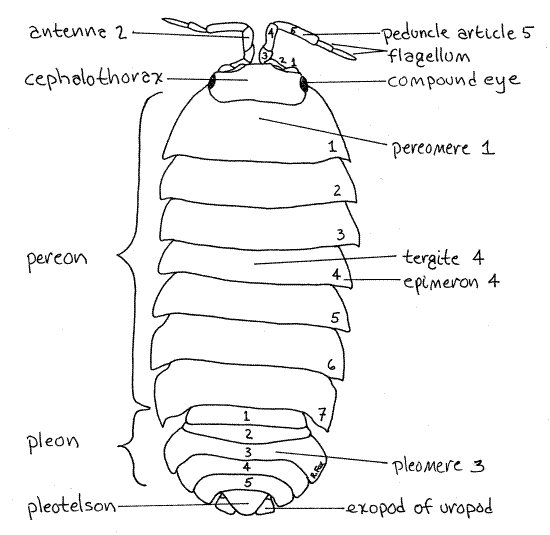
The compound eyes on the sides of the cephalothorax are composed of about 15-50 ommatidia, or facets (Fig 1). The facets are visible with magnification.
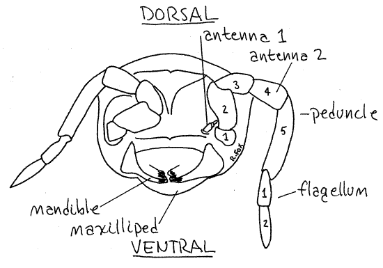
The first two pairs of head appendages are the first and second antennae. The second antennae are large, multiarticulate, filiform structures (Fig 1, 2). Each is composed of a basalpeduncle of five articles and a distal flagellum of a variable number of small articles (Two in Armadillidium and Porcellio, three in Oniscus, and many in Ligia, Fig 6).
In Armadillidium the second antennae fit neatly into broad recesses on the front of the head permitting them to be folded completely out of harm's way when the animal enrolls. The antennae are sensory and, as the animal walks, are used to touch the ground ahead before venturing onto it. This is easily observed in active animals.
The first antennae are tiny and difficult to find until you know exactly where to look (Fig 2). Hold the animal vertically so you can focus on the anterior surface of the head and look medial to the base of the second antennae. The first antennae are between the bases of the second antennae and are composed of only three tiny articles.
The remaining three pairs of head appendages are mouthparts. They are clustered together, along with the maxilliped of the thorax, on the ventral surface of the head, surrounding the mouth. Dissection and study of the mouthparts is difficult in these small animals and your instructor may excuse from attempting it.
The two mandibles are on either side of the mouth. Their incisors and molars are heavily sclerotized. Oniscoidean mandibles lack a palp but a lacinia mobilis is present. The first maxillae are immediately posterior to the mandibles and are followed by the second maxillae.
Posteriorly, the mouthparts are covered and hidden by the maxilliped. This is the fused pair of appendages of the first thoracomere and is the most posterior of the appendages of the cephalothorax. You can see the broad flat maxilliped on the posterior side of the cluster of mouthparts on the ventral head. If you move it posteriorly, you can see the mouthparts.
Figure 2. En face view of the head of Armadillidium. Isopod32L.gif
Pereon
The seven thoracic segments (thoracomeres) independent of the cephalothorax constitute the pereon. Each segment is a pereomere and their appendages are pereopods. The seven pereomeres are easily seen dorsally. The cephalothorax fits into a median notch on the anterior margin of the first pereomere (Fig 1, 19-61).
The pereomeres are good examples of typical arthropod segments. Each is covered by an exoskeletal ring composed of two sclerites. Dorsally is the heavily sclerotized, archedtergite (Fig 1). The seven tergites of the pereon are easy to see and count.
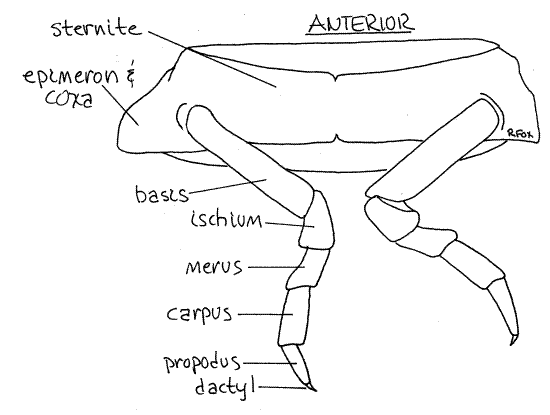
The ventral portion of each segmental exoskeletal ring is the sternite (Fig 3). It is also sclerotized but less so than the tergite. The ventral surface of the pereon is more or less flat although each sternite bears strong ridges and grooves, mostly for reception of the legs when they are folded against the body.
In reproductively active females the sternites are hidden by thin, membranous oostegites, or brood plates. Do not mistake the oostegites for sternites which lie dorsal to them and may be hidden by them. The oostegites are soft and pliable. They will be discussed in more detail later.
If you see white areas on the venter of the anterior pereomeres, it means your specimen is getting ready to molt. These areas are deposits of calcium salts that have been reclaimed from the old cuticle in anticipation of the loss of that cuticle.
The lateral extremities of the tergites reach ventrally far below the level of the sternites. These extensions are the epimera, or side plates (Fig 1, 3). Together the epimera on each side form a wall that encloses and protects the space ventral to the sternites. In females the marsupium occupies this space and in both sexes the legs are here.
The seven pairs of pereopods, or walking legs, resemble each other, a condition responsible for the name "isopod" (iso = equal, pod = foot). Examine one of the pereopods and note that it is uniramous as are the pereopods of most malacostracans (Fig 3). The pereopod arises from the epimeron at its junction with the sternum and epimeron.
Characteristic of malacostracan thoracopods, the leg is composed of a linear series of seven articles. The proximal article is the coxa, which in oniscoideans is fused rigidly with the tergite to form the epimeron. It does not give the appearance of being part of the leg nor is it recognizable as being a distinct part of the epimeron. Consequently the legs appear to be composed of six articles. The first of the six is the long basis which articulates with the coxa of the epimeron (Fig 3). It is followed, in order by the ischium, merus, carpus, and propodus. The final dactyl is small.
Peracarida is characterized by the presence, in breeding females, of a marsupium, or brood pouch, on the ventral pereon of females (Fig 19-52, 19-62). This pouch is formed of thin, membranous brood plates, or oostegites, extending medially from the basis of some of the pereopods. A marsupium is not present in males.
In woodlice the first five pairs of female pereopods bear oostegites. Together these ten flexible plates form the marsupium, into which the eggs are released and in which they develop. The marsupium of terrestrial isopods holds water in which the embryos develop, much as they would in the ancestral aquatic habitat.
>1a. If your specimen is a female, look for the marsupium. If it is present, it will completely cover the ventral aspect of the pereon, obscuring the sternites. If you can see the sternites, there is no marsupium. Each of the oostegites is independent of the others and can be lifted with a nadel. Lift the medial edge of the marsupium and see if eggs, embryos, or juvenile isopods are present. Development in peracarids is direct, without a true larva, and occurs in the marsupium. In most peracarids development produces a miniature replica of the adult but in isopods there is first produced a manca "larva". The manca is also a miniature replica of the adult but has only six pairs of pereopods instead of seven. The seventh pair is added by a subsequent molt.<
Figure 3. Ventral view of pereomere and pereopod 5 of Armadillidium. Isopod33L.gif
In malacostracans the male gonopore is on the eighth thoracic segment, which is pereomere 7 of isopods. In isopods the male gonopore is on a median genital papilla on the ventral surface of this segment (Fig. 4). The papilla is a transparent, membranous triangle, or cone, pointing posteriorly and overlapping the bases of the anterior abdominal appendages.
The female gonopores of malacostracans are on the ventral surface of thoracic segment 6 (pereomere 5) but they are difficult to see. The male gonopods transfer sperm to these openings during copulation.
Abdomen
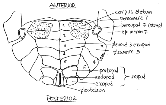
The abdomen is composed of six segments of which the last, or sixth, is fused with the telson to form a pleotelson (Figs 1, 4, 5). The first five segments are independent and together form the pleon (Fig 1). Pleon segments are called pleomeres and their appendages are pleopods. The appendages of the sixth abdominal segment are uropods. The telson, which is the terminal region of the body, is not a true segment.
The biramous pleopods (Figs 4, 5) consist of a basal protopod and two rami. The medial rami, or endopods, are very soft with a thin cuticle and are the gas exchange surfaces, orgills. They are covered and protected by the lateral rami, or exopods, which are more heavily sclerotized and harder (Figs 4, 5). The exopods are moved aside to expose the gills for gas exchange. To see the gills you must look under the exopods.
The long slender endopods (Fig 4) of the first two pleopods of males are modified to serve as copulatory organs, or gonopods . (Only the second endopods of Ligia are so modified.) Sperm transfer is indirect. Sperm from the male gonopore at the tip of the genital papilla is transferred to the female by the gonopods. The pleopods of females are unmodified.
Figure 4. Ventral view of the pleon and pleotelson of a male Armadillidium. Isopod35La.gif
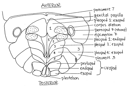
In woodlice the anterior exopods of the pleopods of both sexes contain a respiratory system of pseudotrachea (Figs 4, 5, 19-66B), known as the corpus alatum, to supplement the gills. In living or freshly killed specimens these are easily seen as large, white masses in the outer edges of the anterior pleopod exopods but they are hard to see in preserved specimens.
Pseudotracheae are adaptations for terrestrial existence. Interior gas exchange surfaces such as lungs or trachea are more efficient at conserving water than are gills. Like the trachea of insects, pseudotracheae are tiny tubes extending from an external spiracle into the interior of the appendage (Fig 19-66B). Unlike those of insects, they deliver oxygen to the blood which then distributes it to the tissues. Oniscoidean pseudotracheae and insect tracheae are independent solutions to the problem of terrestrial gas exchange and are an example of convergent evolution.
The uropods are the appendages of the sixth abdominal segment (Fig 1). They are biramous and heavily sclerotized. Each has a basal protopod from which arise an exopod and endopod (Figs 4, 5). In most species the rami are long and visible dorsally extending posteriorly from under the pleotelson. In Armadillidium, however, the rami are short, so they can be included within the sphere when the animal enrolls. The short, flat exopods are the only parts of the uropods visible dorsally (Fig 1). The exopods fit into the gap between the side of the telson and the fifth abdominal tergite. The slender endopods are hidden beneath the pleotelson and can be seen only from the venter (Fig 4, 5). Examine your specimen from the ventral surface and find the three parts of the uropods.
Figure 5. Ventral view of the pleon and pleotelson of a female Armadillidium. Isopod34La.gif
(The exopods of Porcellio and Oniscus are long, pointed, and blade-shaped and extend well posterior to the telson. Their endopods are much shorter and are scarcely visible dorsally. The exopods and endopods of Ligia are similar to each other and both are long and slender.) Theanus opens ventrally under the base of the pleotelson and is hidden by the uropods.
Behavior
>1b. If living unanesthetized Armadillidium are available, stimulate one with an applicator stick and watch it roll into a ball. Inspect the ball with magnification and see if any appendages protrude from it or if there are any gaps in its armor. The chief predators of woodlice are ants, spiders, and shrews, the first two of which are foiled by this behavior. Woodlice also have repugnatorial glands in the epimera and uropods which discourage predators. <
>1c. Under magnification, watch a woodlouse walk and note the movements of the second antennae. Is their behavior consistent with the hypothesis that these appendages are sensory? <
Identification
The most common North American terrestrial isopods associated with human habitations are relatively easy to distinguish at the generic level. The following key and Figure 6 can be used to determine the identity of your specimen.
Figure 6 Distinguishing characteristics of three genera of oniscoideans. A. Oniscus, B. Porcellio, C. Ligia. Redrawn from van Name (1936). Isopod67L.gif
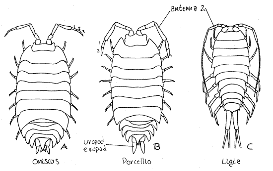
1a. Exopod of uropod extending posteriorly well beyond posterior margin of pleotelson (Fig 6) ..................................................................................................................2
1b. Exopod of uropod extending little, if at all, beyond pleotelson (Fig 1).. Armadillidium
2a. Flagellum of second antenna with more than 10 articles (Fig 6C)….……. Ligia
2b. Flagellum of second antenna with fewer than 5 articles (Fig 6A,B)…………. .3
3a. Flagellum of second antenna with 2 articles (Fig 6B)...…………..….. Porcellio
3b. Flagellum of second antenna with three articles (Fig 6A).……….…… Oniscus
References
Drummond PC . 1965. The terrestrial isopod crustaceans (Oniscoidea) of Florida. MS thesis, University of Florida, Gainesville. 75p.
Sutton SL. 1972. Woodlice. Pergamon, Oxford. 144p.
Ruppert EE, Fox RS, Barnes RB. 2004. Invertebrate Zoology, A functional evolutionary approach, 7 th ed. Brooks Cole Thomson, Belmont CA. 963 pp.
Van Name, W. G. 1936. The American land and fresh-water isopod Crustacea. Bull. American Mus. Nat. Hist. 71:1-535.
Wägele J-W. 1992. Isopoda. in Harrison, F. W. & A. G. Humes (eds.). Microscopic Anatomy of Invertebrates vol. 9 Crustacea. Wiley-Liss, New York.
Supplies
Dissecting microscope
8-cm culture dish
Living or preserved terrestrial isopods
Chloroform
Cotton
Applicator stick